Zombie Aqua™ Fixable Viability Kit

Pricing & Availability
Regulatory Status
RUO
Other Names
Fixable Dye, Fixable Viability Dye, Dead, Apoptosis
Ave. Rating
Submit a Review
Product Citations
publications
b-Zombie_Aqua_Viability_Dye_032713
One day old splenocytes were stained with Zombie Aqua™ and analyzed without fixation (blue) or analyzed after fixation and permeabilization (red). Cells alone without Zombie Aqua™ staining are indicated in black.
  • b-Zombie_Aqua_Viability_Dye_032713
    One day old splenocytes were stained with Zombie Aqua™ and analyzed without fixation (blue) or analyzed after fixation and permeabilization (red). Cells alone without Zombie Aqua™ staining are indicated in black.
  • c-Zombie_Aqua_Viability_Dye_012721
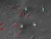
    HeLa cells were treated with 20% EtOH for 20 seconds, washed twice with PBS, and then were left to recover for five minutes with cell culture media in 37°C. The cells were stained with Zombie Aqua™ (1:1000) (cyan) for 15 minutes and then fixed with 1% paraformaldehyde (PFA) for ten minutes. Nuclei were counterstained with DRAQ5 (Red) for five minutes. The image was captured with 40X objective.
See Zombie Aqua™ spectral data
Cat # Size Price Quantity Check Availability Save
423101 100 tests $89
Check Availability


Need larger quantities of this item?
Request Bulk Quote
423102 500 tests $335
Check Availability


Need larger quantities of this item?
Request Bulk Quote
Description

Zombie Aqua™ is an amine-reactive fluorescent dye that is non-permeant to live cells but permeant to cells with compromised membranes. Thus, it can be used to assess live vs. dead status of mammalian cells. Zombie Aqua™ is a polar, water-soluble dye providing very bright green fluorescence, making it suitable for use in multi-color detection.
 

Product Details
Technical data sheet

Product Details

Preparation
Zombie Aqua™ Fixable Viability Kit is composed of lyophilized Zombie Aqua™ dye and anhydrous DMSO. For reconstitution, pre-warm the kit to room temperature; add 100 µl of DMSO to one vial of Zombie Aqua™ dye and mix until fully dissolved. 100 tests = 1 vial of Zombie Aqua™ + DMSO, 500 tests = 5 vials of Zombie Aqua™ + DMSO.
Storage & Handling
Store kit at -20°C upon receipt. Do not open vials until needed. Once the DMSO is added to the Zombie Aqua™ dye, use immediately, or store at -20°C in a dry place and protected from light, preferably in a desiccator or in a container with desiccant for no more than one month.
Application

FC - Quality tested
ICFC, ICC - Verified

Recommended Usage

Each lot of this product is quality control tested by immunofluorescent staining with flow cytometric analysis.

For flow cytometry, the suggested dilution is 1:100-1:1000 for 1-10 million cells. For immunofluorescence microscopy, the suggested dilution is 1:1000. It is recommended that the reagent be titrated for optimal performance for each application, as optimal dosage varies with cell type.


Excitation Laser
Violet Laser (405 nm)
Application Notes

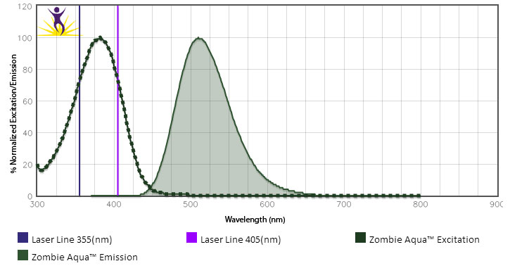
Zombie Aqua™ dye is excited by the violet laser and has a maximum emission of 516 nm. If using in a multi-color panel design, filter optimization may be required depending on other fluorophores used. Zombie Aqua™ dye has similar emission to Brilliant Violet 510™.

Standard Cell Staining Protocol:

  1. Prior to reconstitution, spin down the vial of lyophilized reagent in a microcentrifuge to ensure the reagent is at the bottom of the vial.
  2. For reconstitution, pre-warm the kit to room temperature; add 100 µL of DMSO to one vial of Zombie Aqua™ dye and mix until fully dissolved
  3. Wash cells with PBS buffer (no Tris buffer and protein free).
  4. Dilute Zombie Aqua™ dye at 1:100-1000 in PBS. Resuspend 1-10 x 106 cells in diluted 100 µL Zombie Aqua™ solution. To minimize background staining of live cells, titrate the amount of dye and/or number of cells per 100 µL for optimal performance. Different cell types can have a wide degree of variability in staining based on cell size and degree of cell death.
    • Note: Don’t use Tris buffer as a diluent and be sure that the PBS does not contain any other protein like BSA or FBS.
    • Note: The amount of dye used can also influence the ability to detect apoptotic as well as live and dead cells.
  5. Incubate the cells at room temperature (or 4°C), in the dark, for 15-30 minutes.
  6. Wash one time with 2 mL BioLegend’s Cell Staining Buffer (Cat. No. 420201) or equivalent buffer containing serum or BSA.
  7. Continue performing antibody staining procedure as desired.
  8. Cells can be fixed with paraformaldehyde or methanol prior to permeabilization or can be analyzed without fixation.

No-wash Sequential Staining Protocol:

  1. Wash cells with PBS buffer (no Tris buffer and protein free).
  2. For reconstitution, pre-warm the kit to room temperature; add 100 µL of DMSO to one vial of Zombie Aqua™ dye and mix until fully  dissolved
  3. Determine the total µL volume of antibody cocktail previously titrated and optimized for the assay that will be added to each vial/well of cells based on a final volume of 100 µL. Subtract that antibody volume from the 100 µl total staining volume intended for the assay. In the remaining volume, dilute Zombie Aqua™ dye at 1:100-1000 in PBS as determined by prior optimization at that volume. For example, if you are adding 20 µL of antibody cocktail for a 100 µL total staining volume, use 80 µL of Zombie Aqua™ solution. Resuspend 1-10 x 106 cells in the appropriate volume of Zombie Aqua™ solution. Different cell types can have a wide degree of variability in staining based on cell size and degree of cell death.
    • Note: Don’t use Tris buffer as a diluent and be sure that the PBS does not contain any other protein like BSA or FBS.
    • Note: The amount of dye used can also influence the ability to detect apoptotic as well as live and dead cells.
  4. Incubate for 10-15 minutes at RT (or 4°C), protected from light. Without washing the cells, add the cell surface antibody cocktail and incubate for another 15-20 minutes.
  5. Add 1-2 mL Cell Staining Buffer (Cat. No. 420201) or equivalent buffer containing BSA or serum. Centrifuge to pellet.
  6. Continue with normal fixation and permeabilization procedure. If planning to skip fixation and analyze cells live, complete an additional wash step to minimize any unnecessary background of the live cells.
    • Notes: If the cell type in use cannot tolerate a protein-free environment, then titrate the Zombie Aqua™ dye in the presence of the same amount of BSA/serum as will be present in the antibody staining procedure. A higher amount of Zombie Aqua™ may be required since the BSA/serum will react with and bind up some proportion of the Zombie Aqua™.
Additional Product Notes

View more applications data using this product to validate Veri-Cells™ lyophilized control cells and to study dendritic cell subsets.

Application References

(PubMed link indicates BioLegend citation)
  1. Vom Berg J, et al. 2013. J Exp Med. 210:2803. PubMed
  2. Radovanovic I, et al. 2014. J Immunol. 193:1290. PubMed
  3. Price PJ, et al. 2014. J Virol. 88:10840. PubMed
  4. Schmid MA, et al. 2014. PLoS Pathog. 10:1004541. PubMed
  5. Cohen SB, et al. 2015. J Immunol. 194:210. PubMed
  6. Li J, et al. 2015. Cancer Res. 75:508. PubMed
  7. Price PJ, et al. 2015. J Immunol. 194:1164. PubMed
  8. Michelet X, et al. 2015. J Immunol. 194:2079. PubMed
  9. Tzeng A, et al. 2015. PNAS. 112:3320. PubMed
  10. Peres AG, et al. 2015. Infect Immun. 83:1587. PubMed
  11. Hansmann L, et al. 2015. Cancer Immunol Res. 3:650. PubMed
Product Citations
  1. Zhou R, et al. 2022. EBioMedicine. 75:103762. PubMed
  2. Mo Y, et al. 2022. Front Immunol. 12:799896. PubMed
  3. Luo J, et al. 2022. J Nanobiotechnology. 20:228. PubMed
  4. Castle AR, et al. 2022. PLoS One. 17:e0269342. PubMed
  5. Healy LM, et al. 2020. Glia. 68:811. PubMed
  6. Buchanan MW, et al. 2021. J Orthop Res. 39:1710. PubMed
  7. Bussières-Marmen S, et al. 2017. Cell Mol Immunol. 10.1038/cmi.2016.72. PubMed
  8. Darzianiazizi M, et al. 2020. Int J Mol Sci. 21:00. PubMed
  9. Soon MSF, et al. 2020. Nat Immunol. 1.984027778. PubMed
  10. Lindner SE, et al. 2020. Immunohorizons. 0.356944444. PubMed
  11. Tasker C, et al. 2021. AAPS J. 23:26. PubMed
  12. Lin C, et al. 2020. Cancer Immunol Res. 632:8. PubMed
  13. Kim J, et al. 2021. Front Cell Neurosci. 638098:15. PubMed
  14. Grigsby SM, et al. 2021. Cancers (Basel). 13:. PubMed
  15. Rueschenbaum S, et al. 2021. Front Immunol. 11:581352. PubMed
  16. Fantone K, et al. 2021. Pathogens. 10:. PubMed
  17. Vella JL, et al. 2021. Life Sci Alliance. 4:. PubMed
  18. Nuccitelli R, et al. 2017. J Immunother Cancer . 10.1186/s40425-017-0234-5. PubMed
  19. Kaczanowska S, et al. 2017. Cancer Res. . 10.1158/0008-5472.CAN-17-0653. PubMed
  20. Gálvez-Cancino F, et al. 2018. Oncoimmunology. 7:e1442163. PubMed
  21. Contijoch EJ et al. 2019. eLife. 8 pii: e40553. PubMed
  22. Wu J et al. 2017. Immunity. 47(6):1114-1128 . PubMed
  23. Eiva MA, et al. 2022. Eur J Immunol. 52:96. PubMed
  24. Duraiswamy J, et al. 2021. Cancer Cell. 39:1623. PubMed
  25. Herz J, et al. 2021. Neuron. 109:3609. PubMed
  26. Mills EL, et al. 2022. Cell Metab. 34:140. PubMed
  27. Miek L, et al. 2022. Immunology. 166:47. PubMed
  28. Wolf B, et al. 2022. Immunology. 166:380. PubMed
  29. Iberg CA, et al. 2022. Cell Rep. 39:110657. PubMed
  30. Shcheynikov N, et al. 2022. Hepatology. 76:1248. PubMed
  31. Tang Q, et al. 2022. Mol Ther. 30:2709. PubMed
  32. Adam-Artigues A, et al. 2022. Sci Adv. 8:eabk2746. PubMed
  33. Yang K, et al. 2022. Sci Immunol. 7:eabn2888. PubMed
  34. Sugimoto S, et al. 2022. Nat Metab. 4:775. PubMed
  35. Ma T, et al. 2022. Elife. 11: . PubMed
  36. Benedetti V, et al. 2022. Sci Adv. 8:eabn3986. PubMed
  37. Guasconi L, et al. 2022. J Fungi (Basel). 8: . PubMed
  38. Zhu J, et al. 2022. Research (Wash D C). 2022:9816272. PubMed
  39. Shen Z, et al. 2022. EBioMedicine. 85:104285. PubMed
  40. Martínez-Sabadell A, et al. 2022. Cell Rep. 41:111430. PubMed
  41. Gao Y, et al. 2022. Cell Res. 32:1086. PubMed
  42. Dhar R, et al. 2022. Int J Mol Med. 50: . PubMed
  43. Balood M, et al. 2022. Nature. 611:405. PubMed
  44. Yoo HJ, et al. 2022. Cell Rep. 41:111598. PubMed
  45. Liang Y, et al. 2022. Theranostics. 12:7729. PubMed
  46. Rico-Llanos G, et al. 2022. Front Immunol. 13:1054962. PubMed
  47. Chen W, et al. 2022. Front Immunol. 13:1046426. PubMed
  48. Zia S, et al. 2022. Mol Neurodegener. 17:82. PubMed
  49. Zhuang L, et al. 2022. J Am Heart Assoc. 11:e027228. PubMed
  50. Pleuger C, et al. 2022. Elife. 11: . PubMed
  51. Shen W, et al. 2023. Dis Model Mech. 16: . PubMed
  52. Perveen R, et al. 2023. Hum Cell. 36:583. PubMed
  53. Krammer S, et al. 2022. Cells. 12: . PubMed
  54. Chan L, et al. 2022. Cells. 12: . PubMed
  55. Cousin N, et al. 2022. Cells. 12: . PubMed
  56. Rice CM, et al. 2023. Life Sci Alliance. 6: . PubMed
  57. Zhao Q, et al. 2023. Nutrients. 15: . PubMed
  58. Romine KA, et al. 2023. Leukemia. 37:580. PubMed
  59. Ai L, et al. 2023. Cell Discov. 9:9. PubMed
  60. Gong HH, et al. 2023. Front Immunol. 13:1089064. PubMed
  61. Gonçalves R, et al. 2023. iScience. 26:105972. PubMed
  62. Liu G, et al. 2023. Commun Biol. 6:102. PubMed
  63. Li H, et al. 2023. Front Immunol. 13:1056447. PubMed
  64. Lutz EA, et al. 2022. PNAS Nexus. 1:pgac244. PubMed
  65. Badenes M, et al. 2023. Life Sci Alliance. 6: . PubMed
  66. Pušnik J, et al. 2023. Nat Commun. 14:572. PubMed
  67. Kang J, et al. 2023. JCI Insight. 8: . PubMed
  68. Chan L, et al. 2023. Int J Mol Sci. 24: . PubMed
  69. Babl N, et al. 2023. Front Oncol. 13:1107484. PubMed
  70. Drummer C, et al. 2023. Front Immunol. 14:1113883. PubMed
  71. Li Y, et al. 2022. Brain Behav Immun. 101:1. PubMed
  72. Ryan CB, et al. 2022. Neurobiol Dis. 163:105608. PubMed
  73. Fahrner JE, et al. 2022. Cancer Discov. 12:958. PubMed
  74. Blair TC, et al. 2022. Life Sci Alliance. 5:. PubMed
  75. Ko J, et al. 2022. Nat Biotechnol. 40:1654. PubMed
  76. Zhu Z, et al. 2022. Clin Cancer Res. 28:4820. PubMed
  77. Lutz EA, et al. 2022. Proc Natl Acad Sci U S A. 119:e2205983119. PubMed
  78. Bermea KC, et al. 2022. J Vis Exp. . PubMed
  79. Jia L, et al. 2022. Gut Microbes. 14:2117503. PubMed
  80. Frost JN, et al. 2022. Sci Adv. 8:eabq5384. PubMed
  81. Schuhmacher J, et al. 2022. Cells. 11:. PubMed
  82. Cruz-Reséndiz A, et al. 2022. Front Immunol. 13:1057499. PubMed
  83. Johnson SD, et al. 2022. Front Immunol. 13:1001727. PubMed
  84. Egli J, et al. 2022. MAbs. 14:2143009. PubMed
  85. Lao L, et al. 2023. Cancer Immunol Res. 11:320. PubMed
  86. Cai Z, et al. 2023. Adv Sci (Weinh). 10:e2207155. PubMed
  87. Yang Y, et al. 2023. MBio. 14:e0328522. PubMed
  88. Gao L, et al. 2023. J Nanobiotechnology. 21:56. PubMed
  89. Jun SH, et al. 2023. Nat Commun. 14:982. PubMed
  90. Fernandes S, et al. 2023. iScience. 26:106071. PubMed
  91. Aso K, et al. 2023. Nat Commun. 14:984. PubMed
  92. Medina S, et al. 2023. Water (Basel). 15:. PubMed
  93. Gupta T, et al. 2023. Front Immunol. 14:1044703. PubMed
  94. Xu K, et al. 2023. Elife. 12:. PubMed
  95. Battistello E, et al. 2023. Mol Cell. 83:1216. PubMed
  96. Zhang J, et al. 2023. Front Immunol. 14:1091541. PubMed
  97. Harpur CM, et al. 2023. Clin Transl Immunology. 12:e1443. PubMed
  98. Pankhurst TE, et al. 2023. Cell Rep. 42:112310. PubMed
  99. Ahmed O, et al. 2023. Bioelectron Med. 9:6. PubMed
  100. Freitag PC, et al. 2023. Mol Ther Methods Clin Dev. 29:120. PubMed
  101. Palakurthi B, et al. 2023. Nat Commun. 14:2109. PubMed
  102. Madar J, et al. 2023. Nat Commun. 14:2158. PubMed
  103. Consonni FM, et al. 2023. Front Immunol. 14:1168455. PubMed
  104. Fang Y, et al. 2023. Front Immunol. 14:1162004. PubMed
  105. Chaturvedi S, et al. 2023. Cell. 186:2036. PubMed
  106. Ximerakis M, et al. 2023. Nat Aging. 3:327. PubMed
  107. Liao Y, et al. 2023. ACS Omega. 8:15217. PubMed
  108. Menevse AN, et al. 2023. Acta Neuropathol Commun. 11:75. PubMed
  109. Liu JC, et al. 2023. Int J Mol Sci. 24:. PubMed
  110. Tang L, et al. 2023. Front Immunol. 14:1145441. PubMed
  111. Zhang Z, et al. 2023. Aging Dis. 14:966. PubMed
  112. Malik JR, et al. 2023. Int J Mol Sci. 24:. PubMed
  113. Feng X, et al. 2023. Nat Commun. 14:3208. PubMed
  114. Komoto S, et al. 2023. Cancers (Basel). 15:. PubMed
  115. Vanderleyden I, et al. 2020. Cell Rep. 30:611. PubMed
  116. Lam B, et al. 2021. Cell Rep. 37:109838. PubMed
  117. Ferrere G, et al. 2021. JCI Insight. 6:. PubMed
  118. Chaturvedi S, et al. 2022. Cell. 185:2086. PubMed
  119. Volta V, et al. 2021. Nat Commun. 12:6979. PubMed
  120. Seki SM, et al. 2020. Sci Signal. :13. PubMed
  121. Krausgruber T, et al. 2020. Nature. 583:296. PubMed
  122. Böttcher M, et al. 2018. Oncoimmunology. 7:e1445454. PubMed
  123. Jeevan-Raj B, et al. 2017. Cell Rep. 10.1016/j.celrep.2017.06.071. PubMed
  124. Fujii H, et al. 2015. PLoS One. 10: 0133216. PubMed
  125. Michelet X, et al. 2015. J Immunol. 194:2079. PubMed
  126. Kobayashi T, et al. 2019. Cell. 176:982. PubMed
  127. Kiritsy MC, et al. 2021. Elife. 10:. PubMed
  128. Blake SJ, et al. 2021. Cell Rep Med. 2:100464. PubMed
  129. Lo MW, et al. 2022. Clin Transl Immunology. 11:e1413. PubMed
  130. Lownik JC, et al. 2020. Biochem Biophys Res Commun. 522:442. PubMed
  131. Gálvez-Cancino F, et al. 2017. Vaccine. 10.1016/j.vaccine.2017.06.041. PubMed
  132. Bailey S, et al. 2017. Nat Commun.. 10.1038/s41467-017-01867-9. PubMed
  133. Barry KC, et al. 2018. Nat Med. 24:1178. PubMed
  134. Farsakoglu Y et al. 2019. Cell reports. 26(9):2307-2315 . PubMed
  135. Shin HS, et al. 2022. Sci Rep. 12:17605. PubMed
  136. Meyer Zu Natrup C, et al. 2022. J Clin Invest. Online ahead of print. PubMed
  137. Toomer G, et al. 2022. Viruses. 14:. PubMed
  138. Radovanovic I, et al. 2014. J Immunol. 193:1290. PubMed
  139. Price P, et al. 2014. J Virol. 88:10840. PubMed
  140. Tzeng A, et al. 2015. Proc Natl Acad Sci U S A. 112:3320. PubMed
  141. Yamaguchi M, et al. 2016. J Leukoc Biol. 100: 131 - 141. PubMed
  142. Swee L, et al. 2016. Open Bio. 6: 160293. PubMed
  143. Liu Y, et al. 2022. Cancer Discov. :. PubMed
  144. Imbrechts M, et al. 2022. iScience. 25:104705. PubMed
  145. Munoz MA, et al. 2021. Elife. 10:. PubMed
  146. Gong W, et al. 2021. Oncoimmunology. 10:1997385. PubMed
  147. Gu H, et al. 2021. Immun Inflamm Dis. 9:1686. PubMed
  148. Kremenovic M, et al. 2022. J Immunother Cancer. 10:. PubMed
  149. van der Heide V, et al. 2022. Cell Rep. :110508. PubMed
  150. Witkowski M, et al. 2021. Nature. 600:295. PubMed
  151. Deng X, et al. 2021. Open Biol. 11:200340. PubMed
  152. Yu X, et al. 2021. Nature. 594:560. PubMed
  153. Park JJ, et al. 2021. Nat Commun. 12:1222. PubMed
  154. Kakiyama G, et al. 2020. J Lipid Res. 61:1629. PubMed
  155. Fronik P, et al. 2021. J Med Chem. 64:12132. PubMed
  156. Li LH, et al. 2020. Antiviral Res. 104929:183. PubMed
  157. Jenull S, et al. 2021. Cell Reports. 36(3):109406. PubMed
  158. Gruber T, et al. 2020. JCI Insight. 5:00. PubMed
  159. Kakiyama G, et al. 2020. J Lipid Res. 61:1629. PubMed
  160. Neidleman J, et al. 2020. Elife. 9:00. PubMed
  161. Meehan S, et al. 2019. Commun Biol. 2:229. PubMed
  162. Tan L, et al. 2019. Cell Rep. 27:3657. PubMed
  163. Hartsough EJ, et al. 2019. Cell Death Dis. 10:281. PubMed
  164. Chattopadhyay A, et al. 2018. Sci Rep. 8:9032. PubMed
  165. Martrus G, et al. 2017. PLoS One.. 10.1371/journal.pone.0182532. PubMed
  166. Setten E, et al. 2022. Nat Commun. 13:6499. PubMed
  167. Alberts-Grill N, et al. 2016. J Immunol. 197(12):4651-4662. PubMed
  168. Clement M, et al. 2016. PLoS Pathog. 12:e1006050. PubMed
  169. Torre S, et al. 2016. Nat Immunol. 10.1038/ni.3581. PubMed
  170. Bittner-Eddy P, et al. 2016. J Immunol. 197: 1435 - 1446. PubMed
  171. Berg J, et al. 2013. J Exp Med. 210:2803. PubMed
  172. Mathur S, et al. 2021. JCI Insight. 6:. PubMed
  173. Pedersen JG, et al. 2021. Front Microbiol. 12:763030. PubMed
  174. Almutairi F, et al. 2021. Front Immunol. 12:772288. PubMed
  175. Bresque M, et al. 2022. J Biol Chem. :101711. PubMed
  176. Peng Q, et al. 2022. EBioMedicine. 77:103904. PubMed
  177. Mukherjee D, et al. 2022. Nat Commun. 13:3747. PubMed
  178. Ruetsche D, et al. 2022. Cells. 11:. PubMed
  179. Hagen SH, et al. 2020. Cell Rep. 33:108485. PubMed
  180. Pan H, et al. 2020. Mol Psychiatry. . PubMed
  181. Darzianiazizi M, et al. 2021. Cytokine X. 3:100053. PubMed
  182. Go DM, et al. 2021. Cell Mol Gastroenterol Hepatol. 12:715. PubMed
  183. Wright AKA, et al. 2017. J Allergy Clin Immunol. 140:1430. PubMed
  184. Botuyan MV, et al. 2018. Nat Struct Mol Biol. 25:591. PubMed
  185. Martínez-Sabadell A, et al. 2022. STAR Protoc. 3:101712. PubMed
  186. Tu X, et al. 2022. Nat Commun. 13:6977. PubMed
  187. Peres A, et al. 2015. Infect Immun . 83:1587. PubMed
  188. Yoshida N, et al. 2016. Nat Commun. 7: 12993. PubMed
  189. Fang V, et al. 2016. Nat Immunol. 18:15-25. PubMed
  190. Luo X, et al. 2021. J Biol Chem. 297:101098. PubMed
  191. Johansen AZ, et al. 2022. Pharmaceutics. 14:. PubMed
  192. Rogawski DS, et al. 2021. Nat Commun. 12:2792. PubMed
  193. Cousin N, et al. 2021. Cancer Res. 81:4133. PubMed
  194. Paiva RS, et al. 2021. Eur J Immunol. 51:1968. PubMed
  195. Britton GJ et al. 2019. Immunity. 50(1):212-224 . PubMed
  196. Grozio A, et al. 2019. Nat Metab. 1:47. PubMed
  197. Ramsey HE, et al. 2018. Cancer Discov. 8:1566. PubMed
  198. Parameswaran P, et al. 2017. Cell Host Microbe. 10.1016/j.chom.2017.08.003. PubMed
  199. Chen Z, et al. 2022. Theranostics. 12:6242. PubMed
  200. Tang JJ, et al. 2022. Cell Rep. 39:110987. PubMed
  201. Amberg N, et al. 2016. J Invest Dermatol. 136:2140-2149. PubMed
  202. Cunningham C, et al. 2016. PLoS Pathog. 12: 1005356. PubMed
  203. Schmitt N, et al. 2015. Mol Cancer Ther. 14: 2103-2111. PubMed
  204. Valbuena Perez JV, et al. 2020. Aging Cell. 19:e13156. PubMed
  205. Sellau J, et al. 2020. Nat Commun. 2.860416667. PubMed
  206. Zheng S, et al. 2022. Blood Cancer Discov. 3:103. PubMed
  207. Yang K, et al. 2022. J Clin Invest. 132:. PubMed
  208. Wilson AS, et al. 2022. Nat Commun. 13:528. PubMed
  209. Tang Y, et al. 2021. Cancer Discov. 11:3142. PubMed
  210. Borst L, et al. 2022. Int J Cancer. 150:688. PubMed
  211. Chan JA, et al. 2020. Cell Rep Med. 1:100157. PubMed
  212. Montes de Oca M, et al. 2020. PLoS Pathog. 16:e1008994. PubMed
  213. Jordan PM, et al. 2020. Cell Rep. 33:108247. PubMed
  214. Doty DT, et al. 2020. Int J Mol Sci. 21:00. PubMed
  215. Dillon SM, et al. 2020. Bio Protoc. e3486:10. PubMed
  216. Pantelyushin S, et al. 2021. Cancers (Basel). :13. PubMed
  217. Alsaleh G, et al. 2020. Elife. 9: . PubMed
  218. Stuani L, et al. 2021. J Exp Med. 218:. PubMed
  219. Ceholski D, et al. 2017. Stem Cell Res. 10.1016/j.scr.2017.06.015. PubMed
  220. Metzemaekers M, et al. 2017. Int J Mol Sci. 10.3390/ijms18071513. PubMed
  221. Tien S, et al. 2017. PLoS One. 10.1371/journal.pone.0181338. PubMed
  222. Alvisi G, et al. 2022. Methods Mol Biol. 2559:243. PubMed
  223. Li FJ, et al. 2022. PLoS One. 17:e0277019. PubMed
  224. Denkers S 2015. J Immunol. 195: 2754-2762. PubMed
  225. Garcia-Bates T, et al. 2016. J Immunol. 196: 2870 - 2878. PubMed
  226. Davidson S, et al. 2016. EMBO Mol Med. 8: 1099 - 1112. PubMed
  227. Chakraborty A, et al. 2020. Cell Rep. 32:108179. PubMed
  228. Soldevilla MM, et al. 2019. Mol Ther. 27:1878. PubMed
  229. Romine KA, et al. 2021. Blood Cancer Discov. 2:518. PubMed
  230. Romine KA, et al. 2021. Cancer Discov. . PubMed
  231. Mehta AK, et al. 2021. Nat Cancer. 2:66. PubMed
  232. Li Y, et al. 2021. Brain Behav Immun. 91:267. PubMed
  233. Karagianni AE, et al. 2021. Sci Rep. 11:14292. PubMed
  234. Korangath P, et al. 2020. Bio Protoc. 10:e3822. PubMed
  235. Amanat F, et al. 2021. Cell. 184:3936. PubMed
  236. Acharya A, et al. 2020. J Virol. 95:e01657. PubMed
  237. Audiger C, et al. 2020. J Immunol. 205:121. PubMed
  238. Fernandez-Castaneda A, et al. 2020. Acta Neuropathol. 139:365. PubMed
  239. Köchl R, et al. 2020. Elife. 9:00. PubMed
  240. Honarpisheh P, et al. 2020. J Neuroinflammation. 0.9625. PubMed
  241. Kruse S, et al. 2018. Oncoimmunology. 8:e1524694. PubMed
  242. Louveau A, et al. 2018. Curr Protoc Immunol. 121:1. PubMed
  243. Wilmore JR et al. 2018. Cell host & microbe. 23(3):302-311 . PubMed
  244. Joshi N and Werner S. 2017. PLoS One. 10.1371/journal.pone.0187162. PubMed
  245. Natoli M, et al. 2022. J Immunother Cancer. 10:. PubMed
  246. Wang Z, et al. 2022. Nat Commun. 13:5447. PubMed
  247. OlguÍn-Alor R, et al. 2016. PLoS One. 11:e0167813. PubMed
  248. Crisci E, et al. 2016. J Virol. 90: 4939 - 4950. PubMed
  249. Hansmann L, et al. 2015. Cancer Immunol Res. 3:650. PubMed
  250. Yamazaki T, et al. 2020. Oncoimmunology. 9:1721810. PubMed
  251. Guo J, et al. 2019. Cancer Immunol Res. 1.349305556. PubMed
  252. Reed J, et al. 2020. Bio Protoc. 10:e3607. PubMed
  253. Alikhanyan K, et al. 2021. Cancers (Basel). 13:. PubMed
  254. Hu H, et al. 2021. Cancer Immunol Res. 9:1298. PubMed
  255. Breen EC, et al. 2022. iScience. 25:104488. PubMed
  256. Zhang F, et al. 2020. EMBO Mol Med. e12034:12. PubMed
  257. Xing J, et al. 2021. Nat Commun. 12:2681. PubMed
  258. Hildebrand KM, et al. 2021. PLoS One. 16:e0253864. PubMed
  259. Littwitz-Salomon E, et al. 2021. Nat Commun. 12:5376. PubMed
  260. Kallert S, et al. 2017. Nature Communications. 10.1038/ncomms15327. PubMed
  261. Rechtien A, et al. 2017. Cell Rep. 10.1016/j.celrep.2017.08.023. PubMed
  262. Yang Y, et al. 2016. Nucleic Acids Res. 44: 4174 - 4188. PubMed
  263. Coria L, et al. 2016. J Immunol. 196: 4014 - 4029. PubMed
  264. Carmona SJ, et al. 2020. Oncoimmunology. 9:1737369. PubMed
  265. , et al. 2021. Eur J Immunol. 51:2708. PubMed
  266. Luo J, et al. 2022. J Nanobiotechnology. 20:228. PubMed
  267. Shen J, et al. 2022. Front Immunol. 13:762580. PubMed
  268. Momin N, et al. 2022. Nat Commun. 13:109. PubMed
  269. Giesen D, et al. 2020. Clin Cancer Res. 26:3999. PubMed
  270. Tsang M, et al. 2020. Am J Pathol. 190:206. PubMed
  271. Dieterich LC, et al. 2020. Angiogenesis. 1.24375. PubMed
  272. Brugat T, et al. 2017. Nat Microbiol. 2:16276. PubMed
  273. Haloul M, et al. 2019. Sci Rep. 9:14050. PubMed
  274. Engelbertsen D, et al. 2019. Sci Rep. 9:10608. PubMed
  275. Freire DM, et al. 2019. Mol Cell. 73:1282. PubMed
  276. Scarneo SA, et al. 2022. Sci Rep. 12:18091. PubMed
  277. Ren Y, et al. 2022. J Immunother Cancer. 10:. PubMed
  278. Andriessen E, et al. 2016. EMBO Mol Med. 8(12):1366-1379. PubMed
  279. Vidy A, et al. 2016. PLoS One. 11:e0165361. PubMed
  280. Krämer B, et al. 2016. PLoS One. 11: 0162068. PubMed
  281. Schmid M, et al. 2016. PLoS One. 12: 1005676. PubMed
  282. Contreras F, et al. 2016. J Immunol. 196: 4143 - 4149. PubMed
  283. Larmonier C, et al. 2016. J Biol Chem. 291: 8918-8930. PubMed
  284. Lim X, et al. 2016. Proc Natl Acad Sci U S A. 113: 1498 - 1505. PubMed
  285. Alikhanyan K, et al. 2020. Immun Inflamm Dis. 8:181. PubMed
  286. Zebertavage LK, et al. 2020. Sci Rep. 10:7376. PubMed
  287. Bourque J, et al. 2021. Heliyon. 7:e08311. PubMed
  288. Alameh MG, et al. 2021. Immunity. 54:2877. PubMed
  289. Bankova AK, et al. 2022. Transplant Cell Ther. . PubMed
  290. Sinha SR, et al. 2020. Cell Host Microbe. 27:659. PubMed
  291. Bullard BL, et al. 2022. NPJ Vaccines. 7:65. PubMed
  292. Rossignol J, et al. 2022. iScience. 25:104353. PubMed
  293. Karagianni A, et al. 2022. Front Oncol. 12:929498. PubMed
  294. Castro JT, et al. 2022. Nat Commun. 13:4831. PubMed
  295. Coden ME, et al. 2020. J Immunol. 204:438. PubMed
  296. James KR, et al. 2020. Nat Immunol. 1.113194444. PubMed
  297. Lewandowski JP, et al. 2020. Genome Biol. 1.039583333. PubMed
  298. Sabree SA, et al. 2021. J Immunother Cancer. 9: . PubMed
  299. Fu Z, et al. 2021. Nat Commun. 12:4462. PubMed
  300. Pan W, et al. 2021. J Immunol. 206:1719. PubMed
  301. Rhys HI, et al. 2018. EBioMedicine. 29:60. PubMed
  302. Fragniere AMC, et al. 2019. Sci Rep. 9:2288. PubMed
  303. Kemp V, et al. 2018. Cancer Gene Ther. 26:268. PubMed
  304. Neal JT, et al. 2018. Cell. 175:1972. PubMed
  305. Cohen S, et al. 2015. J Immunol. 194:210. PubMed
  306. Price P, et al. 2015. J Immunol. 194:1164. PubMed
  307. Chattopadhyay A, et al. 2016. J Lipid Res. 57: 832 - 847. PubMed
  308. Dutzan N, et al. 2017. Immunity. 46(1):133-147. PubMed
  309. Xiao L, et al. 2021. Clin Cancer Res. 27:4338. PubMed
  310. Rocha-Resende C, et al. 2021. J Mol Cell Cardiol. 157:98. PubMed
  311. Pathania AS, et al. 2022. Mol Ther Oncolytics. 25:308. PubMed
  312. Barington L, et al. 2022. Cells. 11:. PubMed
  313. Bendtsen SK, et al. 2022. Oncoimmunology. 11:2026020. PubMed
  314. Jiang Y, et al. 2021. Cell Death Differ. 28:3009. PubMed
  315. Huo Y, et al. 2021. BMC Genomics. 22:145. PubMed
  316. Madsen A, et al. 2020. Immunity. 53:852. PubMed
  317. Li H, et al. 2021. Adv Sci (Weinh). 2001596:8. PubMed
  318. Ritzel RM, et al. 2021. Glia. 69:746. PubMed
  319. Parayath NN, et al. 2020. Nat Commun. 4.680555556. PubMed
  320. Wang L, et al. 2020. EMBO J. 39:e104514. PubMed
  321. Tron AE, et al. 2018. Nat Commun. 9:5341. PubMed
  322. Collin R, et al. 2017. J Immunol. 10.4049/jimmunol.1600789. PubMed
  323. Teng Y, et al. 2017. J Hematol Oncol. 10.1186/s13045-017-0485-0. PubMed
  324. Huo Y, et al. 2022. Front Immunol. 13:983116. PubMed
  325. Clark GC, et al. 2022. Front Oncol. 12:913656. PubMed
  326. Ryan P, et al. 2016. Proc Natl Acad Sci U S A. 113(50):14378-14383. PubMed
  327. LM S, et al. 2016. Cell Rep. 16(12): 3286-96. PubMed
  328. Miller JE, et al. 2021. JCI Insight. 6:. PubMed
  329. Wen T, et al. 2022. Cancer Immunol Res. 10:162. PubMed
  330. Lee GJ, et al. 2022. Oncoimmunology. 11:2015170. PubMed
  331. Andriessen EMMA, et al. 2021. EMBO Mol Med. 13:e11754. PubMed
  332. Shibata H, et al. 2021. Oncoimmunology. 10:1958589. PubMed
  333. Constant DA, et al. 2022. Immunohorizons. 6:416. PubMed
  334. Li J, et al. 2022. Nat Commun. 13:4032. PubMed
  335. Taylor D, et al. 2022. Front Immunol. 13:936129. PubMed
  336. Yoder A, et al. 2017. PLoS Pathog. 13(2):e1006226. PubMed
  337. Davey M, et al. 2017. Nat Commun. 10.1038/ncomms14760. PubMed
  338. Fang H, et al. 2020. Nat Commun. 4.661805556. PubMed
  339. Hagen SH, et al. 2021. STAR Protocols. 2(3):100641. PubMed
  340. Daemen S, et al. 2021. STAR Protoc. 2:100511. PubMed
  341. Santos NB, et al. 2021. PLoS Pathog. 17:e1009381. PubMed
  342. Madsen D, et al. 2017. Cell Rep.. 10.1016/j.celrep.2017.12.011. PubMed
  343. Becker JR, et al. 2018. Nat Commun. 9:5406. PubMed
  344. Suan D et al. 2017. Immunity. 47(6):1142-1153 . PubMed
  345. Schmid M, Harris E 2014. PLoS Pathog. 10:1004541. PubMed
  346. Sakamoto K, et al. 2021. Immunity. 54:2321. PubMed
  347. McCallion A, et al. 2022. Front Immunol. 13:961599. PubMed
  348. Ye Q, et al. 2022. NPJ Vaccines. 7:84. PubMed
  349. Alice AF, et al. 2021. Sci Rep. 11:16347. PubMed
  350. Singh AK, et al. 2022. Nat Commun. 13:878. PubMed
  351. Kumar S, et al. 2022. Gut. . PubMed
  352. Bent EH, et al. 2021. Nat Commun. 12:6218. PubMed
  353. Manouchehri N, et al. 2021. Proc Natl Acad Sci U S A. 118:. PubMed
  354. Nylund P, et al. 2021. Cell Death Dis. 12:167. PubMed
  355. Hofmann J, et al. 2021. Front Immunol. 11:599495. PubMed
  356. Kakiyama G, et al. 2020. J Lipid Res. 61:1629. PubMed
  357. de Winde CM, et al. 2021. J Cell Sci. 134: . PubMed
  358. Xueyang Yu et al. 2017. Immunity. 47(5):903-912 . PubMed
  359. Ogrinc Wagner A, et al. 2019. PLoS One. 14:e0210998. PubMed
  360. Zwick M, et al. 2019. Front Immunol. 10:222. PubMed
  361. Li H, et al. 2022. Front Immunol. 13:946202. PubMed
  362. Morikawa M, et al. 2016. PLoS One. 11:e0163607. PubMed
  363. Concha-Benavente F, et al. 2016. Cancer Res . 76: 1031 - 1043. PubMed
  364. Arenas EJ, et al. 2021. Nat Commun. 12:1237. PubMed
  365. Rodriguez E, et al. 2021. Cancers (Basel). 13:. PubMed
  366. Dholakia J, et al. 2022. Gynecol Oncol. 164:170. PubMed
  367. Wegrecki M, et al. 2022. Nat Commun. 13:3872. PubMed
  368. Peng Z, et al. 2022. J Neuroinflammation. 19:186. PubMed
  369. Brockman QR, et al. 2022. JCI Insight. :. PubMed
  370. Lee SH, et al. 2022. Nat Commun. 13:5461. PubMed
  371. Lu YJ, et al. 2021. Cell Rep. 36:109696. PubMed
  372. Schuldt N, et al. 2017. The Journal of Immunology. 10.4049/jimmunol.1700406. PubMed
  373. Pardo E, et al. 2017. PLoS One. 12(6):e0177472. PubMed
  374. Dawson NA, et al. 2019. JCI Insight. 4:6. PubMed
  375. Vigeland CL, et al. 2019. Physiol Rep. 7:e14019. PubMed
  376. Leong Y, et al. 2016. Nat Immunol. 17:1187-1196. PubMed
  377. Ohs I, et al. 2016. Nat Commun. 7:13708. PubMed
  378. Jerby–Arnon L, et al. 2018. Cell. 175:984. PubMed
  379. Pfirschke C, et al. 2020. Cell Rep. 32:108164. PubMed
  380. Ghezraoui H, et al. 2018. Nature. 560:122. PubMed
  381. Reuschl AK, et al. 2022. Cell Rep. 39:110650. PubMed
  382. Mello AM, et al. 2022. Oncogenesis. 11:56. PubMed
  383. Leary N, et al. 2022. J Extracell Vesicles. 11:e12197. PubMed
  384. Li M, et al. 2021. Mol Biomed. 2:41. PubMed
  385. Milich LM, et al. 2021. J Exp Med. 218:. PubMed
  386. Alhudaithi SS, et al. 2020. Mol Pharm. 17:4691. PubMed
  387. Fucà G, et al. 2021. J Immunother Cancer. 9:. PubMed
  388. Egedal JH, et al. 2021. Mucosal Immunol. 14:1203. PubMed
  389. Casselli T, et al. 2021. PLoS Pathog. 17:e1009256. PubMed
  390. Abdelsamed HA, et al. 2020. Nat Immunol. 1.276388889. PubMed
  391. Rocha-Resende C, et al. 2020. JCI Insight. 5:00. PubMed
  392. Luo X, et al. 2020. J Clin Invest. 130:1635. PubMed
  393. Peter Morawski, Chen-Feng Qi, Silvia Boll 2017. Sci Rep. 7:40838. PubMed
  394. Antsiferova M, et al. 2017. EMBO Mol Med. 9(1):27-45. PubMed
  395. Ju X, et al. 2016. J Immunol. 197(12):4613-4625. PubMed
  396. Sebina I, et al. 2016. PLoS Pathog. 12:e1005999. PubMed
  397. Gouwy M, et al. 2016. PLoS One. 11:e0166006. PubMed
  398. McGuire V, et al. 2016. Sci Rep. 6:31159. PubMed
  399. Comte D, et al. 2016. Proc Natl Acad Sci U S A. 113: 9321 - 9326. PubMed
  400. Carpenter S, et al. 2016. PLoS Pathog. 12: 1005380. PubMed
  401. Dhimolea E, et al. 2021. Cancer Cell. 39:240. PubMed
  402. Li Y, et al. 2022. Theranostics. 12:5364. PubMed
  403. Burger ML, et al. 2021. Cell. 184:4996. PubMed
  404. Patel C, et al. 2022. Curr Protoc. 2:e400. PubMed
  405. Neidleman J, et al. 2017. PLoS Pathog. 13(2):e1006163. PubMed
  406. Sam J, et al. 2020. Front Oncol. 10:575737. PubMed
  407. Yang C, et al. 2020. Cell Host Microbe. 467:27. PubMed
  408. Blair TC, et al. 2020. J Immunol. 204:3416. PubMed
  409. Martini E, et al. 2016. Cell Rep. 14:1062-1073. PubMed
  410. Weiss G, et al. 2016. Mucosal Immunol. 10.1038/mi.2016.104. PubMed
  411. Li Y, et al. 2020. Theranostics. 10:11376. PubMed
  412. Bozoglu T, et al. 2022. Adv Sci (Weinh). 9:e2103867. PubMed
  413. Lau P, et al. 2022. Cell Mol Immunol. :. PubMed
  414. van Loon K, et al. 2022. Cancers (Basel). 14:. PubMed
  415. Singh SS, et al. 2022. Clin Transl Immunology. 11:e1396. PubMed
  416. Sakamoto K, et al. 2022. STAR Protoc. 3:101052. PubMed
  417. Verma M, et al. 2021. J Exp Med. 218:. PubMed
  418. Van Winkle JA, et al. 2020. J Virol. 94:. PubMed
  419. Liu X, et al. 2021. Adv Sci (Weinh). 8:e2100233. PubMed
  420. Kohn M, et al. 2021. Front Immunol. 626627:12. PubMed
  421. Salahudeen AA, et al. 2020. bioRxiv. . PubMed
  422. Tomlinson JE, et al. 2018. J Neuroinflammation. 0.753472222. PubMed
  423. Padhye A, et al. 2019. Sci Rep. 9:4819. PubMed
  424. Yuan C, et al. 2018. iScience. 11:13. PubMed
  425. Tiberti S, et al. 2022. Nat Commun. 13:6752. PubMed
  426. Farooq F, et al. 2016. Sci Rep. 6:27944. PubMed
  427. Pflügler S, et al. 2020. Commun Biol. 3:252. PubMed
  428. Luo ZW, et al. 2021. Int J Nanomedicine. 16:2949. PubMed
  429. Xu L, et al. 2022. Front Immunol. 13:841141. PubMed
  430. Spath S, et al. 2022. iScience. 25:104998. PubMed
  431. Cho SX, et al. 2020. Nat Commun. 4.481944444. PubMed
  432. Filippo Buono M, et al. 2020. Cells. 9:00. PubMed
  433. Friedrich V, et al. 2021. Gut Microbes. 13:1. PubMed
  434. Jiang M, et al. 2021. Front Pharmacol. 12:657486. PubMed
  435. Henkle TR, et al. 2021. Cancer Res. 81:4560. PubMed
  436. Schaller MA, et al. 2021. JCI Insight. 6:e148003. PubMed
  437. Pan L, et al. 2021. Stem Cell Res Ther. 12:496. PubMed
  438. Dai H, et al. 2017. J Immunol. 10.4049/jimmunol.1700418. PubMed
  439. Krogmann A, et al. 2016. PLoS One. 11: 0146326. PubMed
  440. Johns S, et al. 2016. Circ Res . 119: 210 - 221. PubMed
  441. Binnewies M, et al. 2019. Cell. 177:556. PubMed
  442. Wang H, et al. 2022. J Cancer. 13:2126. PubMed
  443. Bouchard G, et al. 2022. Cancer Res. 82:648. PubMed
  444. Khurana P, et al. 2021. Front Immunol. 12:700374. PubMed
  445. Lal JC, et al. 2021. Breast Cancer Res. 23:83. PubMed
  446. Chakraborty A, et al. 2021. Biomaterials. 273:120796. PubMed
  447. Feng Y, et al. 2022. EClinicalMedicine. 43:101226. PubMed
  448. Tseng SH, et al. 2021. J Biomed Sci. 28:63. PubMed
  449. Tee YC, et al. 2021. FASEB Bioadv. 3:829. PubMed
  450. Xia L, et al. 2021. Cancer Immunol Res. 9:707. PubMed
  451. Chiou SH, et al. 2021. Immunity. 54:586. PubMed
  452. Schreurs RRCE, et al. 2021. Mucosal Immunology. 14(3):605-614. PubMed
  453. Workel HH, et al. 2020. Int J Mol Sci. 21:00. PubMed
  454. Laczkó D, et al. 2020. Immunity. 53:724. PubMed
  455. Salahudeen AA, et al. 2020. Nature. 588:670. PubMed
  456. Orozco SL, et al. 2019. Cell Rep. 28:2275. PubMed
  457. Kimura K, et al. 2018. Nat Commun. 9:17. PubMed
  458. Wang CJH, et al. 2022. J Biol Chem. :102714. PubMed
  459. Scheyltjens I, et al. 2022. Nat Protoc. 17:2354. PubMed
  460. Hatfield J, Brown M 2015. Cell Immunol. . PubMed
  461. Li J, et al. 2015. Cancer Res. 75:508. PubMed

Antigen Details

Biology Area
Apoptosis/Tumor Suppressors/Cell Death, Cell Biology, Neuroscience
Gene ID
NA

Related FAQs

Is Zombie Aqua™ an equivalent to cell labeling and tracer dyes such as CellTrace™ Violet and CFSE?

Zombie Aqua™ is not a direct equivalent to such dyes because their mechanism of action and scope of application are entirely different.

I am concerned about the spillover I am observing from the Zombie dye into its neighboring channels.
Rule of thumb with Zombie dyes is to titrate them down as much as possible to fit your application. This should potentially help with spillover. Secondly, Zombie positive events represent dead cells and are typically gated out from analysis.
How does the performance of your Zombie dye compare with competitors?

Zombie dyes have been tested against other leading competitors' fixable viability kits and given comparable results. We also highly recommend that you titrate down the amount of each dye used in order to best match the negative signals of your unstained sample and MFI- (mean fluorescence intensity) stained samples.

Can I use methanol/ethanol for fixation after using a Zombie dye?

Yes, most fixation reagents are fine to be used with Zombie dyes. However, it should be noted that Zombie dyes can still be sensitive to reactive oxygen species. Light exposure or reagents with hydrogen peroxide can lead to free radical formation, affecting fluorescence.

Can Zombie be used to determine bacteria, yeast viability?
We have not tested in house bacterial or yeast viability using Zombie dyes. It is not clear whether the difference between surface and intracellular signals will be significantly different in case of non mammalian cells.
Can I use Zombie with cells suspension containing serum?
Serum is full of proteins which will sequester the dye and thereby reducing its effective concentration. The basic rule of thumb with zombie is to titrate it based on your specific condition. Titration also helps reduce the background and spillover into other channels.
Can I use Zombie dyes for microscopy?

Zombie dyes tested in-house for microscopy applications will display data on the product technical datasheet. It should be noted that Zombie dyes may not work for dead cell discrimination in every microscopy application. Important considerations that may impact analysis are determining the signal level that constitutes a dead cell and identifying the proper plane to observe the dead cells.

Why can't I fix my cells prior to using Zombie dyes?

The fixation process can contort and alter the membrane of cells, effectively rendering them dead. Since the ability of the Zombie dyes to stain dead cells is correlated with cell permeability, your results may no longer be a valid representation of dead versus live cells.

Can I use Zombie dyes to detect apoptotic cells?

Yes, Zombie dyes can be used with apoptosis markers, such as Annexin V or Apotracker™ (shown below), to discriminate live, apoptotic, and dead cells.

One day-old C57BL/6 mouse thymocytes were stained with Apotracker™ Tetra Alexa Fluor® 647 and Zombie™ YG581. Zombie-dim/Apotracker™-positive cells are apoptotic, while double-positive cells are dead. Live cells are negative for both markers.

Can I use the UV laser to stimulate Zombie Aqua™? If so, can I then use it in conjunction with BV510™?

While we typically do not test Zombie Aqua™ with the UV laser, its excitation peak suggests it is effectively excited at 355 nm. However, we would not recommend using BV510™ off the violet laser and Zombie Aqua™ off the UV laser at the same time. Due to cross-beam excitation of BV510™ by the UV laser and the violet excitation of Zombie Aqua™, this would lead to significantly increased background and excessive compensation requirements.

How should I store Zombie dyes?

Store the Zombie dye kit at -20°C upon receipt. Do not open vials until needed. Once DMSO is added, use immediately or store at -20°C in a dry place and protected from light, preferably in a desiccator or in a container with desiccant for no more than one month.

Go To Top Version: 10    Revision Date: 10/16/2025

For Research Use Only. Not for diagnostic or therapeutic use.


This product is supplied subject to the terms and conditions, including the limited license, located at www.biolegend.com/terms) ("Terms") and may be used only as provided in the Terms. Without limiting the foregoing, BioLegend products may not be used for any Commercial Purpose as defined in the Terms, resold in any form, used in manufacturing, or reverse engineered, sequenced, or otherwise studied or used to learn its design or composition without express written approval of BioLegend. Regardless of the information given in this document, user is solely responsible for determining any license requirements necessary for user’s intended use and assumes all risk and liability arising from use of the product. BioLegend is not responsible for patent infringement or any other risks or liabilities whatsoever resulting from the use of its products.


BioLegend, the BioLegend logo, and all other trademarks are property of BioLegend, Inc. or their respective owners, and all rights are reserved.


8999 BioLegend Way, San Diego, CA 92121 www.biolegend.com
Toll-Free Phone: 1-877-Bio-Legend (246-5343) Phone: (858) 768-5800 Fax: (877) 455-9587

ProductsHere

Login/Register
Remember me
Forgot your password? Reset Password
Request an Account