FITC Annexin V

Pricing & Availability
Regulatory Status
RUO
Other Names
Annexin A5, Dead, Apoptosis
Ave. Rating
Submit a Review
Product Citations
publications
AnnexinV_FITC_092908.jpg
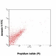
Human T leukemia cell line, Jurkat, non-treated (top) or treated (bottom) with BioLegend’s anti-human CD95 (EOS9.1) mAb (cat. 305704) for 6 hours, then stained with Annexin V-FITC and Propidium Iodide (PI) (cat. 421301)
  • AnnexinV_FITC_092908.jpg
    Human T leukemia cell line, Jurkat, non-treated (top) or treated (bottom) with BioLegend’s anti-human CD95 (EOS9.1) mAb (cat. 305704) for 6 hours, then stained with Annexin V-FITC and Propidium Iodide (PI) (cat. 421301)
  • B_AnnexinV_FITC_092908.jpg
See FITC spectral data
Cat # Size Price Quantity Check Availability Save
640905 25 tests 67€
Check Availability


Need larger quantities of this item?
Request Bulk Quote
640906 100 tests 156€
Check Availability


Need larger quantities of this item?
Request Bulk Quote
640945 300 tests 324€
Check Availability


Need larger quantities of this item?
Request Bulk Quote
Description

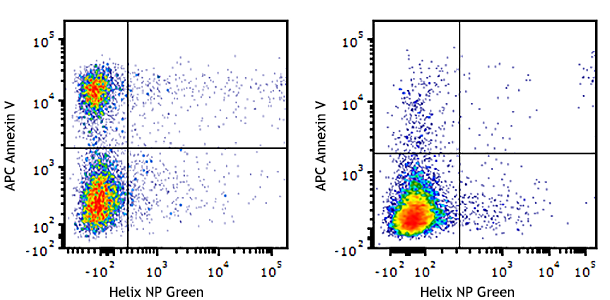
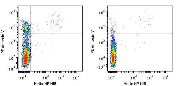
Annexin V (or Annexin A5) is a member of the annexin family of intracellular proteins that binds to phosphatidylserine (PS) in a calcium-dependent manner. PS is normally only found on the intracellular leaflet of the plasma membrane in healthy cells, but during early apoptosis, membrane asymmetry is lost and PS translocates to the external leaflet. Fluorochrome-labeled Annexin V can then be used to specifically target and identify apoptotic cells. Annexin V Binding Buffer (cat. no. 422201) is recommended for use with Annexin V staining.Annexin V binding alone cannot differentiate between apoptotic cells and necrotic. Therefore, we recommend using our Helix NP™ Blue (Cat. No. 425305), Helix NP™ Green (Cat.  No. 425303) or Helix NP™ NIR (Cat. No. 425301). Early apoptotic cells will exclude 7-AAD and PI, while late stage apoptotic cells and necrotic cells will stain positively, due to the passage of these dyes into the nucleus where they bind to DNA.

Product Details
Technical Data Sheet (pdf)

Product Details

Verified Reactivity
Human, Mouse, Rat
Reported Reactivity
Other Species
Formulation
Phosphate-buffered solution, pH 7.2, containing 0.09% sodium azide and BSA (origin USA)
Preparation
The purified protein was conjugated with FITC under optimal conditions.
Concentration
Lot-specific (to obtain lot-specific concentration and expiration, please enter the lot number in our Certificate of Analysis online tool.)
Storage & Handling
The Annexin V solution should be stored undiluted between 2°C and 8°C, and protected from prolonged exposure to light. Do not freeze.
Application

FC - Quality tested

Recommended Usage

Each lot of this product is quality control tested by immunofluorescent staining with flow cytometric analysis. For flow cytometric staining, the suggested use of this reagent is 5 µl per 100,000 - million cells in a 100 µl volume of Annexin V Binding Buffer (Cat No. 422201). It is recommended that the reagent be titrated for optimal performance for each application.

Excitation Laser
Blue Laser (488 nm)
Application Notes

Annexin V Staining

  1. Wash cells twice with cold BioLegend Cell Staining Buffer (Cat. No. 420201) and then resuspend cells in Annexin V Binding Buffer (Cat. No. 422201) at a concentration of 1x10⁶ cells/mL.
  2. Transfer 100 µL of cell suspension in 5 mL test tube.
  3. Add 5 µL of fluorochrome conjugated Annexin V.
  4. Stain with a viability dye, such as PI (Cat. No. 421301), 7-AAD (Cat. Nos. 420403 & 420404), or Helix NP dyes (Cat. Nos. 425301, 425303, & 425305), if desired.
  5. Gently vortex the cells and incubate for 15 min at RT (25°C) in the dark.
  6. Add 400* µL of Annexin V Binding Buffer (Cat. No. 422201) to each tube. *For more concentrated samples, add a minimum of 200 µl of Annexin V Binding Buffer in this step.
  7. Analyze by flow cytometry.

For a better experience detecting apoptosis, we now recommend Apotracker™. Cell staining with Apotracker™ is Calcium independent. Thus, no special buffers are required, and the protocol can be shortened for single-step co-staining with other reagents.

Application References
  1. Koopman G, et al. 1994. Blood 84:1415.
  2. Vermes I, et al. 1995. J. Immunol. Methods 184:39.
  3. Dachary-Prigent J, et al. 1993. Blood 81:2554.
  4. Sekine C, et al. 2009. Int Immunol. PubMed
  5. Grujic M, et al. 2010. J. Immunol. 185:1730. PubMed
  6. Mulik RS, et al. 2010. Int J Pharm. 398:190. PubMed
  7. Gupta A, et al. 2011. Leuk Res. 35:1498. PubMed
  8. Speth C,et al. 2013. J Infect Dis. 207:823. PubMed
  9. Wang Q, et al. 2013. Genes Dev. 27:615. PubMed
  10. Gill K, et al. 2013. Biocheim Biophys Acta. 830:2763. PubMed
  11. Juel HB, et al. 2013. PLoS One. 8:64619. PubMed
  12. Hellmann J, et al. 2013. J. Immunol. 191:1383. PubMed
  13. Sequier S, et al. 2013. PLoS One. 8:70937. PubMed
  14. Galski H, et al. 2013. Biochem Pharmacol. 86:584. PubMed
  15. Chen YL, et al. 2013. J Exp Med. PubMed
  16. Lin WH, et al. 2014. PLoS One. 9:83160. PubMed
  17. Salge-Bartles U, 2014. Thromb res. 133:244. PubMed
  18. Zhang X, et al. 2014. Acta Biochim Biophys Sin. 46:261. PubMed
  19. Intermann J, et al. 2014. Cell Signal. 26:1567. PubMed
  20. de Vires M, 2014. Am J Physiol Lung Cell Mol Physiol. 307:240. PubMed
  21. M Parrula MC, et al. 2014. Virus Res. 189:206. PubMed
  22. Passaro C, et al. 2014. Mol Oncol. 9:78. PubMed
  23. Tsou WI, et al. 2014. J Biol Chem. 289:25750. PubMed
  24. Awojoodu AO, et al. 2014. Blood. 124:1941. PubMed
  25. Xiong R, et al. 2014. Toxicol Appl Pharmacol. 280:285. PubMed
  26. Liu J, et al. 2015. J Mol Cell Cardiol. 80:10. PubMed
Product Citations
  1. Derudder E, et al. 2016. Proc Natl Acad Sci U S A. 113: 5065-5070. PubMed
  2. Uriostegui–Arcos M, et al. 2020. Open Biol. 10:200050. PubMed
  3. Yao Z, et al. 2018. J Virol. 92:00:00. PubMed
  4. Glynn RA, et al. 2022. J Immunol. 208:371. PubMed
  5. Cao H, et al. 2022. Br J Haematol. 198:155. PubMed
  6. Ugamraj HS, et al. 2022. MAbs. 14:2095949. PubMed
  7. Cao L, et al. 2022. Cell Rep. 40:111156. PubMed
  8. Choi UY, et al. 2022. Cell Rep. 40:111234. PubMed
  9. Kimmerling RJ, et al. 2022. Commun Biol. 5:1295. PubMed
  10. Chen X, et al. 2022. Front Pharmacol. 13:1028692. PubMed
  11. Shahid AM, et al. 2022. PLoS One. 17:e0278209. PubMed
  12. Önel T, et al. 2023. Naunyn Schmiedebergs Arch Pharmacol. :. PubMed
  13. Papakonstantinou A, et al. 2022. Int J Mol Sci. 24: . PubMed
  14. Li J, et al. 2023. J Biol Chem. 299:102875. PubMed
  15. Martinelli M, et al. 2023. iScience. 26:105860. PubMed
  16. Tay HG, et al. 2023. Mol Ther. 31:825. PubMed
  17. Dean BD, et al. 2023. J Immunother Cancer. 11: . PubMed
  18. Lieschke E, et al. 2022. Cell Death Differ. 29:1004. PubMed
  19. Gallego-Murillo JS, et al. 2022. Biotechnol Bioeng. 119:3096. PubMed
  20. Du X, et al. 2023. Nucleic Acids Res. 51:1050. PubMed
  21. Vanoli F, et al. 2023. Oncogenesis. 12:8. PubMed
  22. Lei K, et al. 2023. iScience. 26:106001. PubMed
  23. Ohuchi K, et al. 2023. Biomed Rep. 18:28. PubMed
  24. Do-Thi VA, et al. 2023. Cancer Res Commun. 3:80. PubMed
  25. Perez GI, et al. 2023. Vaccines (Basel). 11:. PubMed
  26. Segaliny AI, et al. 2023. Commun Biol. 6:380. PubMed
  27. Potez M, et al. 2023. Front Oncol. 13:1124272. PubMed
  28. Woodley K, et al. 2023. Nat Commun. 14:2132. PubMed
  29. Quintela M, et al. 2023. Clin Epigenetics. 15:63. PubMed
  30. Quintela M, et al. 2023. Br J Cancer. . PubMed
  31. Zelenka T, et al. 2022. Nat Commun. 13:6954. PubMed
  32. Zhang T, et al. 2022. EBioMedicine. 77:103872. PubMed
  33. Dhoonmoon A, et al. 2022. Nat Commun. 13:5063. PubMed
  34. Kitayama J, et al. 2016. PLoS One. 11: 0154542. PubMed
  35. Gedye C, et al. 2016. Sci Rep. 6: 25220. PubMed
  36. Carlsson J, et al. 2015. PLoS One. 10: 0143741. PubMed
  37. Bouhaddou M, et al. 2018. PLoS Comput Biol. 14:e1005985. PubMed
  38. Espinosa-Cueto P, et al. 2017. PLoS One.. 10.1371/journal.pone.0182126. PubMed
  39. Dowling CM, et al. 2021. Sci Adv. 7:. PubMed
  40. Lin YN, et al. 2022. Oncoimmunology. 11:2027136. PubMed
  41. Kim EH, et al. 2020. Elife. 9:00. PubMed
  42. Gupta A, et al. 2011. Leuk Res. 35:1498. PubMed
  43. Fujii T, et al. 2015. Proc Natl Acad Sci U S A. 112: 12800 - 12805. PubMed
  44. Cardoso B, et al. 2015. PLoS One. 10: 0143897. PubMed
  45. Yamauchi T et al. 2018. Cancer cell. 33(3):386-400 . PubMed
  46. Feng W, et al. 2017. Nat Commun. 0.697916667. PubMed
  47. Jones PD, et al. 2018. Arterioscler Thromb Vasc Biol. 38:1711. PubMed
  48. Olelewe C, et al. 2022. iScience. 25:104340. PubMed
  49. Wang Q, et al. 2022. Nat Commun. 13:3022. PubMed
  50. Alcon C, et al. 2020. Cell Death Dis. 0.898611111. PubMed
  51. Figueiredo T, et al. 2020. Mol Psychiatry. . PubMed
  52. Lopes-Coelho F, et al. 2021. Front Oncol. 11:656229. PubMed
  53. Bowen ME, et al. 2019. Dev Cell. 50:212. PubMed
  54. Sales–Dias J, et al. 2019. PLoS One. 14:e0217002. PubMed
  55. Santos LC, et al. 2019. Nat Commun. 1.328472222. PubMed
  56. Lim JA, et al. 2018. BMC Cancer. 0.860416667. PubMed
  57. Venter C, et al. 2017. Cell Physiol Biochem. 43:1288. PubMed
  58. Kim Y, et al. 2020. J Neurochem. 155:177. PubMed
  59. Hassan MIA, et al. 2021. Comput Struct Biotechnol J. 19:880. PubMed
  60. Ramalingam P, et al. 2020. J Exp Med. 217:00:00. PubMed
  61. Soprano M, et al. 2016. PLoS One. 11: 0155970. PubMed
  62. Fortin J, et al. 2020. Cancer Cell. 37:308. PubMed
  63. Karavasili C, et al. 2017. International Journal of Pharmaceutics. 10.1016/j.ijpharm.2017.05.061. PubMed
  64. Thompson PJ et al. 2019. Cell metabolism. 29(5):1045-1060 . PubMed
  65. Noguchi F, et al. 2017. Mol Cancer Ther. 1.316666667. PubMed
  66. Kumar S, et al. 2018. Cancers (Basel). 0.724305556. PubMed
  67. Coray M, et al. 2022. Int J Mol Sci. 23:. PubMed
  68. Vereertbrugghen A, et al. 2021. Front Oncol. 11:598319. PubMed
  69. Sun V, et al. 2021. Front Immunol. 12:716661. PubMed
  70. Macpherson L, et al. 2020. Nature. 577:266. PubMed
  71. Pfalzgraff A, et al. 2016. Sci Rep. 6:31577. PubMed
  72. O'Neill K, et al. 2016. Genes Dev. 30: 973-988. PubMed
  73. Liu J, et al. 2015. J Mol Cell Cardiol. 80:10. PubMed
  74. Vries M, et al. 2014. Am J Physiol Lung Cell Mol Physiol. 307:240. PubMed
  75. Chen MS, et al. 2019. Mol Cancer Res. 17:697. PubMed
  76. Ni F, et al. 2019. Cell Stem Cell. 24:608. PubMed
  77. Ning X, et al. 2019. Mol Cell. 74:19. PubMed
  78. Greenblatt SM, et al. 2018. Cancer Cell. 33:1111. PubMed
  79. Naqvi N,et al. 2017. Sci Rep.. 10.1038/s41598-017-12753-1. PubMed
  80. Wang M, et al. 2017. Biochemical and Biophysical Research Communications. 10.1016/j.bbrc.2017.05.097. PubMed
  81. Beeravolu N, McKee C, Chaudhry G 2017. J App Toxicol. 10.1002/jat.3469. PubMed
  82. Kim OV, et al. 2022. iScience. 25:104654. PubMed
  83. Shahzadi SK, et al. 2022. Clin Sci (Lond). 136:181. PubMed
  84. Kolbrink B, et al. 2022. Biochem J. 479:259. PubMed
  85. He C, et al. 2021. Oncoimmunology. 1897295:10. PubMed
  86. Parrula M, et al. 2014. Virus Res. 189:206. PubMed
  87. Brigger D, et al. 2015. Cell Death Dis. 6: e1861. PubMed
  88. Zhang J, et al. 2016. Cell Death Dis. 7:e2266. PubMed
  89. Wang D, et al. 2016. J Cell Sci. 129: 2261 - 2272. PubMed
  90. Uchtenhagen H, et al. 2016. Nat Commun. 7:12614. PubMed
  91. Geiger R, et al. 2016. Cell. 167:829-842. PubMed
  92. Esteghamat F, et al. 2019. Nat Genet. 51:1233. PubMed
  93. Blümel E, et al. 2020. Oncoimmunology. 9:1751561. PubMed
  94. Sekine C, et al. 2009. Int Immunol. 1.079861111. PubMed
  95. Kinsella S, et al. 2021. Cell Rep. 37:109789. PubMed
  96. O'Connor MJ, et al. 2021. Oncogene. 40:4872. PubMed
  97. Rauzan M, et al. 2017. PLoS One. 12:e0174107. PubMed
  98. Liu Y, et al. 2017. Acta Biomater. 10.1016/j.actbio.2017.08.021. PubMed
  99. Chiani M, et al. 2017. Artif Cells Nanomed Biotechnol. 10.1080/21691401.2017.1337029. PubMed
  100. Yang L, et al. 2021. Cell Death Differ. 28:2616. PubMed
  101. Martín Monreal MT, et al. 2021. Front Immunol. 12:716250. PubMed
  102. Schmitz RL, et al. 2021. Cells. 10:. PubMed
  103. Babaei A, et al. 2021. Adv Pharm Bull. 11:361. PubMed
  104. Madala HR, et al. 2020. Cancer Res. 3492:80. PubMed
  105. Clements KE, et al. 2020. Nat Commun. 4.706944444. PubMed
  106. Salge-Bartels U, et al. 2014. Thromb Res. 133:244. PubMed
  107. Passaro C, et al. 2014. Mol Oncol. 9:78. PubMed
  108. Ligtenberg M, et al. 2016. J Immunol. 196: 759 - 766. PubMed
  109. Hrdinka M, et al. 2016. PLoS One. 11: 0162863. PubMed
  110. Blümel E, et al. 2019. Oncoimmunology. 8:e1641387. PubMed
  111. Kamiyama N, et al. 2017. Antiviral Res. 10.1016/j.antiviral.2017.08.007. PubMed
  112. Montero–Herradón S, et al. 2017. Organogenesis. 0.675. PubMed
  113. Nunes SC, et al. 2018. Sci Rep. 6.939583333. PubMed
  114. Sharif T, et al. 2018. Autophagy. 1.101388889. PubMed
  115. Eichelmann AK, et al. 2018. Sci Rep. 8:17553. PubMed
  116. Zhu Y, et al. 2022. Clin Transl Med. 12:e887. PubMed
  117. Khatib JB, et al. 2022. Oncotarget. 13:1078. PubMed
  118. Krauss PL, et al. 2021. Front Immunol. 12:730672. PubMed
  119. Herold N, et al. 2017. Nat Med. 23:256-263. PubMed
  120. Xie S, et al. 2021. Sci Rep. 0.660416667. PubMed
  121. Müller I, et al. 2020. Mol Cell. 970:77. PubMed
  122. Morimoto A, et al. 2021. Nat Commun. 12:2136. PubMed
  123. Norkin M, et al. 2021. Cell Rep. 35:109026. PubMed
  124. Yoon Y, et al. 2016. Stem Cell Reports. 7:840-853. PubMed
  125. Chheda Z, et al. 2016. J Immunol. 197: 2016 - 2026. PubMed
  126. Slone E, et al. 2015. J Leukoc Biol. 98: 791 - 804. PubMed
  127. Séguier S, et al. 2013. PLoS One. 8:70937. PubMed
  128. Liu H, et al. 2020. Cell Death Dis. 0.767361111. PubMed
  129. Zervou S, et al. 2017. PLoS One.. 10.1371/journal.pone.0182994. PubMed
  130. Ishidome T et al. 2017. EBioMedicine. 22:89-99 . PubMed
  131. Matsuda K, et al. 2021. Cell Rep Methods. 1:100122. PubMed
  132. Vanoli F, et al. 2021. Oncogene. 40:5095. PubMed
  133. Liu H, et al. 2021. J Immunother Cancer. 9:. PubMed
  134. Smith CM, et al. 2021. Biochem J. 478:3331. PubMed
  135. Li Y, et al. 2021. Sci Transl Med. 13:. PubMed
  136. Meyer N, et al. 2021. J Clin Med. 10:. PubMed
  137. Douguet L, et al. 2016. PLoS One. 11:e0166164. PubMed
  138. Toutounchian J, et al. 2016. Mol Pharmacol. 91(1):1-13. PubMed
  139. Grujic M, et al. 2010. J Immunol. 185:1730. PubMed
  140. Wang Q, et al. 2013. Genes Dev. 27:615. PubMed
  141. Litzenburger UM, et al. 2017. Genome Biol. 18:15. PubMed
  142. Rajavel T, et al. 2017. Sci Rep. 2.665277778. PubMed
  143. Meyer N, et al. 2021. Int J Mol Sci. 22:. PubMed
  144. Kolbrink B, et al. 2022. Cell Mol Life Sci. 79:387. PubMed
  145. Pajarillo E, et al. 2020. J Biol Chem. 295:3040. PubMed
  146. Gholamrezaei M, et al. 2021. Iran J Parasitol. 15:475. PubMed
  147. L Chang, Y Li, D Kaplan 2016. Sci Rep. 6:36862. PubMed
  148. Becker A, et al. 2016. Sci Rep. 6: 33847. PubMed
  149. Kumai T, et al. 2015. Sci Rep. 5:16280. PubMed
  150. Polak R, et al. 2015. Blood. 126: 2404 - 2414. PubMed
  151. Matsumoto A, et al. 2019. J Extracell Vesicles. 9:1696517. PubMed
  152. Pirali T, et al. 2017. ChemMedChem. 1.570833333. PubMed
  153. Ishida M, et al. 2021. Int J Mol Sci. 22:. PubMed
  154. Lee DH, et al. 2020. Circ Res. 127:1182. PubMed
  155. Alsina-Sanchis E, et al. 2020. Mol Cancer Res. . PubMed
  156. Galski H, et al. 2013. Biochem Pharmacol. 86:584. PubMed
  157. Tsou W, et al. 2014. J Biol Chem. 289:25750. PubMed
  158. Putheti P, et al. 2010. PLoS One. 5:e8706. PubMed
  159. Hafez A, et al. 2017. Oncogenesis. 10.1038/oncsis.2017.46. PubMed
  160. Erdogan M, et al. 2022. Chem Biodivers. 19:e202200659. PubMed
  161. Molinar-Inglis O, et al. 2021. Proc Natl Acad Sci U S A. 118:. PubMed
  162. Dhoonmoon A, et al. 2020. Nucleic Acids Res. 48:7252. PubMed
  163. Nastasi C, et al. 2021. Sci Rep. 11:1458. PubMed
  164. Liu B, et al. 2016. J Biol Chem. 291: 23869 - 23881. PubMed
  165. Lhuillier C, et al. 2015. J Biol Chem. 290: 16797 - 16811. PubMed
  166. Passaro C, et al. 2013. Endocr Relat Cancer. 20:633. PubMed
  167. Mulik R, et al. 2010. Int J Pharm. 398:190. PubMed
  168. Speth C, et al. 2013. J Infect Dis. 207:823. PubMed
  169. Gladow N, et al. 2020. PLoS One. 15:e0227734. PubMed
  170. Lee HW, et al. 2018. Am J Physiol Renal Physiol. 314:F280. PubMed
  171. Ki H, et al. 2017. Biomed Pharmacother. 10.1016/j.biopha.2017.09.118. PubMed
  172. Shadboorestan A, et al. 2022. Iran J Basic Med Sci. 25:506. PubMed
  173. Huang Y, et al. 2022. Front Cell Dev Biol. 9:837777. PubMed
  174. Babaei A, et al. 2020. Daru. 28:555. PubMed
  175. King R, et al. 2021. Sci Adv. 7:eabj5293. PubMed
  176. Cai Z, et al. 2020. Cell Rep. 31:107816. PubMed
  177. Lin W, et al. 2014. PLoS One. 9:83160. PubMed
  178. Zhang X, et al. 2014. Acta Biochim Biophys Sin. 46:261. PubMed
  179. CO L, et al. 2015. Nat Commun. 6: 7739. PubMed
  180. Di Somma S, et al. 2019. Cancers (Basel). 1.70625. PubMed
  181. Schuler F, et al. 2017. Nat Commun. . 10.1038/s41467-017-01850-4. PubMed
  182. Jackson LM, et al. 2021. Nucleic Acids Res. 49:12855. PubMed
  183. Huang YL, et al. 2020. Elife. 9:00. PubMed
  184. Abu El Maaty MA, et al. 2021. Sci Adv. 7: . PubMed
  185. Bahl S, et al. 2021. Cell Death Dis. 12:469. PubMed
  186. Jones P, et al. 2016. J Biol Chem. 291: 16318 - 16327. PubMed
  187. Awojoodu A, et al. 2014. Blood. 124:1941. PubMed
  188. Intemann J, et al. 2014. Cell Signal. 26:1567. PubMed
  189. Labi V, et al. 2019. Genes Dev. 33:1673. PubMed
  190. Fang P, et al. 2019. Commun Biol. 0.315972222. PubMed
  191. Kumar S, et al. 2019. Int J Cancer. 146:1652. PubMed
  192. Zhao CC, et al. 2018. Oncol Rep. 40:3392. PubMed
  193. Schleicher EM, et al. 2018. Nucleic Acids Res. 46:8908. PubMed
  194. Thomas A, et al. 2017. PLoS One.. 10.1371/journal.pone.0183542. PubMed
  195. Tatangelo V, et al. 2022. Front Oncol. 12:877495. PubMed
  196. Alcon C, et al. 2021. Cells. 10:. PubMed
  197. Cao H, et al. 2021. Nat Commun. 12:1792. PubMed
  198. Faria M, et al. 2017. PLoS One. 12(2):e0172689. PubMed
  199. Clements KE, et al. 2018. Nucleic Acids Res. 46:8898. PubMed
  200. Monzen S, et al. 2022. Mol Clin Oncol. 16:19. PubMed
  201. Konda P, et al. 2022. Am J Cancer Res. 12:210. PubMed
  202. Wu J, et al. 2021. Adv Sci (Weinh). 8:e2101029. PubMed
  203. Bostanabad SY, et al. 2021. Sci Rep. 11:1539. PubMed
  204. Coutaz M, et al. 2016. Sci Rep. 6:39117. PubMed
  205. Akane K, et al. 2016. Proc Natl Acad Sci U S A. 113: 2460 - 2465. PubMed
  206. Xiong R, et al. 2014. Toxicol Appl Pharmacol. 280:285. PubMed
  207. Chen Y, et al. 2013. J Exp Med. 210:2515. PubMed
  208. Mendès C, et al. 2019. Sci Rep. 9:14107. PubMed
  209. Lopes–Coelho F, et al. 2017. Oncotarget. 8:82803. PubMed
  210. Lu Y, et al. 2018. Cancer Cell. 33:1048. PubMed
  211. Thanuthanakhun N, et al. 2017. PLoS One. . 10.1371/journal.pone.0187610. PubMed
  212. Kuntz E, et al. 2017. Nat Med. 10.1038/nm.4399. PubMed
  213. Tu Z, et al. 2022. Cell Rep. 40:111072. PubMed
  214. Bensberg M, et al. 2021. Proc Natl Acad Sci U S A. 118:. PubMed
  215. Paparella AS, et al. 2021. Nat Commun. 12:6285. PubMed
  216. Huang YL, et al. 2021. iScience. 24:103168. PubMed
  217. Zhang Y, et al. 2021. Cell Death Dis. 0.563194444. PubMed
  218. Kang JH, et al. 2020. Cell Death Dis. 0.803472222. PubMed
  219. Dewdney B, et al. 2020. Sci Rep. 10:16769. PubMed
  220. Simoni L, et al. 2020. Cell Rep. 33:108330. PubMed
  221. Lyu X, et al. 2019. BMC Genomics. 1.235416667. PubMed
  222. Jing Y, et al. 2019. J Allergy Clin Immunol. 144:1377. PubMed
  223. Quan M, et al. 2019. Oncol Rep. 41:3051. PubMed
  224. Siddhartha V, et al. 2017. Artif Cells Nanomed Biotechnol . 10.1080/21691401.2017.1313267. PubMed
  225. He J, et al. 2017. FASEB J. 31:2893. PubMed
  226. Kasikara C, et al. 2021. J Clin Invest. 131:. PubMed
  227. Ning N, et al. 2022. Theranostics. 12:5537. PubMed
  228. Bottardi S, et al. 2020. Exp Hematol. 68:88. PubMed
  229. Langsch S, et al. 2016. Cancer Res . 76: 4160 - 4169. PubMed
  230. Xiong R, et al. 2015. Mol Pharmacol. 88: 1045 - 1054. PubMed
  231. Hellmann J, et al. 2013. J Immunol. 191:1383. PubMed
  232. Juel H, et al. 2013. PLoS One. 8:64619. PubMed
  233. Gill K, et al. 2013. Biochim Biophys Acta. 1830:2763. PubMed
  234. Young MM, et al. 2019. Cell Death Dis. 1.004861111. PubMed
  235. Santos I, et al. 2019. Nutrients. 2.210416667. PubMed
  236. Vancsik T, et al. 2019. Cancer Med. 3.313888889. PubMed
  237. Zheng M, et al. 2019. Proc Natl Acad Sci U S A. 116:12422. PubMed
  238. Zheng Q, et al. 2017. Mol Oncol. 10.1002/1878-0261.12056. PubMed
  239. Wang Y, et al. 2022. Front Cardiovasc Med. 9:876087. PubMed
  240. Shankman LS, et al. 2021. Curr Biol. 31:2469. PubMed
  241. Du Z, et al. 2020. J Allergy Clin Immunol. . PubMed
  242. Pahari S, et al. 2020. Autophagy. 1.375694444. PubMed
  243. Schleicher EM, et al. 2020. PLoS Genet. 16:e1009176. PubMed
RRID
AB_2561291 (BioLegend Cat. No. 640905)
AB_2561292 (BioLegend Cat. No. 640906)
AB_2629519 (BioLegend Cat. No. 640945)

Antigen Details

Biology Area
Apoptosis/Tumor Suppressors/Cell Death, Cell Biology, Neuroscience
Gene ID
308 View all products for this Gene ID
Specificity (DOES NOT SHOW ON TDS):
Annexin V
Specificity Alt (DOES NOT SHOW ON TDS):
Annexin V
App Abbreviation (DOES NOT SHOW ON TDS):
FC
Hidden Names (DOES NOT SHOW ON TDS):
A5, AnV, ANX5, ENX2, PP4, Apoptosis, Dead
UniProt
View information about Annexin V on UniProt.org

Related FAQs

How is your Annexin made and what sequence does it cover?

It is made in E. coli, covering human aa Met1-Asp320.

How does pH and staining temperature affect Annexin V-Phosphatidylserine binding?

Annexin-Phosphatidylserine binding is lost below pH 5.2 and with prolonged incubation over a temperature of 42°C.

Why do I need to use Annexin V Binding Buffer?

Annexin V binding requires the presence of calcium in the solution.  So, we provide Annexin V Binding Buffer (cat # 422201), which is optimized for the best performance of Annexin V staining.

Can I use RPMI during Annexin V staining?

It is best to follow protocol as described on the product data sheet. Moreover, RPMI 1640 has a relatively high concentration of phosphate and low calcium ion concentration, which negatively impacts Annexin binding to its target phosphatidylserine (PS). Measurement of cell death by using Annexin V may also be significantly affected by time of incubation on ice, calcium concentration, and type of medium.

Can I freeze Annexin V conjugates?

It should not be frozen as it will lead to loss of biological activity due to dimerization.

Is Annexin V suitable for conjugation with the Maxpar® kit for CyTOF®?

Maxpar® Labeling kits require the protein to be partially reduced, so the metal chelate can be introduced through an SH group in the hinge region of the reduced antibody. Human Annexin V contains only one Cysteine which was reported to be chemically inactive. Thus, the Maxpar® labeling protocol would not work with Annexin V, unless a free –SH group can be introduced to Annexin V.  For more information regarding SH-mediated conjugation of Annexin V please consult published papers such as this one.

Go To Top Version: 7    Revision Date: 10/22/2024

For Research Use Only. Not for diagnostic or therapeutic use.


This product is supplied subject to the terms and conditions, including the limited license, located at www.biolegend.com/terms) ("Terms") and may be used only as provided in the Terms. Without limiting the foregoing, BioLegend products may not be used for any Commercial Purpose as defined in the Terms, resold in any form, used in manufacturing, or reverse engineered, sequenced, or otherwise studied or used to learn its design or composition without express written approval of BioLegend. Regardless of the information given in this document, user is solely responsible for determining any license requirements necessary for user’s intended use and assumes all risk and liability arising from use of the product. BioLegend is not responsible for patent infringement or any other risks or liabilities whatsoever resulting from the use of its products.


BioLegend, the BioLegend logo, and all other trademarks are property of BioLegend, Inc. or their respective owners, and all rights are reserved.


8999 BioLegend Way, San Diego, CA 92121 www.biolegend.com
Toll-Free Phone: 1-877-Bio-Legend (246-5343) Phone: (858) 768-5800 Fax: (877) 455-9587

This data display is provided for general comparisons between formats.
Your actual data may vary due to variations in samples, target cells, instruments and their settings, staining conditions, and other factors.
If you need assistance with selecting the best format contact our expert technical support team.

ProductsHere

Login / Register
Remember me
Forgot your password? Reset password?
Create an Account