


 United States ($) USD
 Austria (€) EUR
 Belgium (€) EUR
 Denmark (DKK) DKK
 Finland (€) EUR
 France (€) EUR
 Germany (€) EUR
 Ireland (€) EUR
 Japan (¥) JPY
 Luxembourg (€) EUR
 Netherlands (€) EUR
 Norway (NOK) NOK
 Sweden (SEK) SEK
 Switzerland (CHF) CHF
 United Kingdom (£) GBP

- Clone
- MP6-XT22 (See other available formats)
- Regulatory Status
- RUO
- Other Names
- Tumor necrosis factor-α, Cachectin, Necrosin, Macrophage cytotoxic factor (MCF), Differentiation inducing factor (DIF), TNFSF-2, TNF-a, TNF-alpha
- Isotype
- Rat IgG1, κ
- Ave. Rating
- Submit a Review
- Product Citations
- publications
-

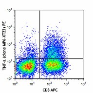
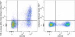
PMA + Ionomycin-stimulated C57BL/6 mouse splenocytes (in the presence of monensin) were stained with CD3 APC, fixed, permeabilized and then stained with TNF-α (clone MP6-XT22) PE (top) or rat IgG1, κ PE isotype control (bottom). -

| Cat # | Size | Price | Save |
|---|---|---|---|
| 506305 | 25 µg | ¥12,110 | |
| 506306 | 100 µg | ¥42,370 |
TNF-α is secreted by macrophages, monocytes, neutrophils, T-cells, and NK-cells. Many transformed cell lines also secrete TNF-α. Monomeric mouse TNF-α is a 156 amino acid protein (N-glycosylated) with a reported molecular weight of 17.5 kD. TNF-α forms multimeric complexes; stable trimers are most common in solution. A 26 kD membrane form of TNF-α has also been described. TNF-α binding to surface receptors elicits a wide array of biologic activities including: cytolysis and cytostasis of many tumor cell lines in vitro, hemorrhagic necrosis of tumors in vivo, increased fibroblast proliferation, and enhanced chemotaxis and phagocytosis in neutrophils.
Product DetailsProduct Details
- Verified Reactivity
- Mouse
- Antibody Type
- Monoclonal
- Host Species
- Rat
- Immunogen
- E. coli-expressed, recombinant mouse TNF-α
- Formulation
- Phosphate-buffered solution, pH 7.2, containing 0.09% sodium azide.
- Preparation
- The antibody was purified by affinity chromatography, and conjugated with PE under optimal conditions.
- Concentration
- 0.2 mg/ml
- Storage & Handling
- The antibody solution should be stored undiluted between 2°C and 8°C, and protected from prolonged exposure to light. Do not freeze.
- Application
-
ICFC - Quality tested
- Recommended Usage
-
Each lot of this antibody is quality control tested by intracellular immunofluorescent staining with flow cytometric analysis. For flow cytometric staining, the suggested use of this reagent is ≤ 0.25 µg per 106 cells in 100 µl volume. It is recommended that the reagent be titrated for optimal performance for each application.
- Excitation Laser
-
Blue Laser (488 nm)
Green Laser (532 nm)/Yellow-Green Laser (561 nm)
- Application Notes
-
ELISA or ELISPOT Detection: The biotinylated MP6-XT22 antibody is useful as a detection antibody for a sandwich ELISA or ELISPOT assay, when used in conjunction with purified 6B8 antibody (Cat. Nos. 510802 & 510804) as the capture antibody.
ELISA Capture: The purified MP6-XT22 antibody is useful as the capture antibody in a sandwich ELISA when used in conjunction with the biotinylated Poly5160 antibody (Cat. No. 516003) as the detection antibody and recombinant mouse TNF-α (Cat. No. 575209) as the standard.
Flow Cytometry6,11,12:The fluorochrome-labeled MP6-XT22 antibody is useful for intracellular immunofluorescent staining and flow cytometric analysis to identify TNF-a-producing cells within mixed cell populations.
Neutralization1,5,10,16,17:The MP6-XT22 antibody can neutralize the bioactivity of natural or recombinant TNF-α. The LEAF™ purified antibody (Endotoxin < 0.1 EU/µg, Azide-Free, 0.2 µm filtered) is recommended for neutralization of mouse TNF-α bioactivity in vivo and in vitro(Cat. No. 506310). For in vivo studies or highly sensitive assays, we recommend Ultra-LEAF™ purified antibody (Cat. No. 506332) with a lower endotoxin limit than standard LEAF™ purified antibodies (Endotoxin < 0.01 EU/µg).
Additional reported applications (for the relevant formats) include: Western blotting, immunohistochemical staining of paraformaldehyde-fixed, saponin-treated frozen tissue sections7-9 in vivo detection5, immunofluorescence, and immunocytochemistry.
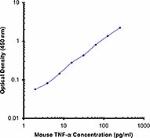
Note: For testing mouse TNF-α in serum, plasma or supernatant, BioLegend's ELISA Max™ Sets (Cat. No. 430901) are specially developed and recommended. -
Application References
(PubMed link indicates BioLegend citation) -
- Abrams J, et al. 1992. Immunol. Rev. 127:5. (Neut)
- Abrams J, et al. 1995. Curr. Prot. Immunol. John Wiley and Sons, New York. Unit 6.20
- Mo X, et al. 1995. J. Virol. 69:1288.
- Sarawar S, et al. 1994. J. Immunol. 153:1246.
- Via C, et al. 2001. J. Immunol. 167:6821. (Neut)
- Infante-Duarte C, et al. 2000 J. Immunol. 165:6107. (FC)
- Jacobs M, et al. 2000. Immunology 100:494. (IHC)
- Marinova-Mutachieva L, et al. 1997. Clin. Exp. Immunol. 107:507. (IHC)
- Williams RO, et al. 2000. J. Immunol. 165:7240. (IHC)
- Scanga CA, et al. 1999. Infect. Immun. 67:4531. (Neut)
- Akilov OE, et al. 2007. J. Leukoc. Biol. 2007;10.1189/jlb.0706439. (FC)
- Lawson BR, et al. 2007. J. Immunol. 178:5366. (FC)
- Patole PS, et al. 2005. J. Am. Soc. Nephrol. 16:3273. PubMed
- Wu S, et al. 2005. Neurosci Lett. 394:158. PubMed
- Carlson MJ, et al. 2009. Blood 113:1365. PubMed
- Shivakumar P, et al. 2017. JCI Insight. 2:e88747 1. PubMed
- Kearney CJ, et al. 2017. Cell Death Differ. 10.1038/cdd.2017.94. PubMed
- Product Citations
-
- RRID
-
AB_315426 (BioLegend Cat. No. 506305)
AB_315427 (BioLegend Cat. No. 506306)
Antigen Details
- Structure
- TNF superfamily; dimer/trimer; 17.5-150 kD (Mammalian)
- Bioactivity
- Paracrine/endocrine mediator of inflammatory and immune functions; selectively cytotoxic for transformed cells; endothelial cell alterations; chemoattractant
- Cell Sources
- Activated monocytes, neutrophils, macrophages, T cells, B cells, NK cells, LAK cells
- Cell Targets
- Monocytes, neutrophils, macrophages, T cells, fibroblasts, endothelial cells, osteoclasts, adipocytes, astroglia, microglia
- Receptors
- TNFRSF1A (TNF-R1, CD120a, TNFR-p60 Type β, p55); TNFRSF1B (TNF-R2, CD120b, TNFR-p80 Type A, p75)
- Cell Type
- Tregs
- Biology Area
- Immunology, Innate Immunity
- Molecular Family
- Cytokines/Chemokines
- Antigen References
-
1. Fitzgerald K, et al. Eds. 2001. The Cytokine FactsBook. Academic Press, San Diego.
2. Beutler B, et al. 1988. Annu. Rev. Biochem. 57:505.
3. Beutler B, et al. 1989. Annu. Rev. Immunol. 7:625.
4. Tracey K, et al. 1993. Crit. Care Med. 21:S415. - Regulation
- Processed by TACE for secretion; upregulated by interferons, IL-2, GM-CSF, substance P, bradykinin, PAF, immune complexes, and cyclooxygenase; downregulated by IL-6, TGF-β, vitamin D3, prostaglandin E2, and PAF antagonists
- Gene ID
- 21926 View all products for this Gene ID
- UniProt
- View information about TNF-alpha on UniProt.org
Related Pages & Pathways
Pathways


Related FAQs
- What type of PE do you use in your conjugates?
- We use R-PE in our conjugates.
Other Formats
View All TNF-α Reagents Request Custom ConjugationCustomers Also Purchased
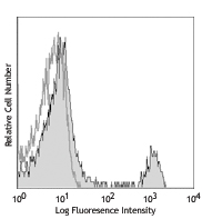
Compare Data Across All Formats
This data display is provided for general comparisons between formats.
Your actual data may vary due to variations in samples, target cells, instruments and their settings, staining conditions, and other factors.
If you need assistance with selecting the best format contact our expert technical support team.
-
Brilliant Violet 785™ anti-mouse TNF-α
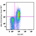
PMA + Ionomycin-stimulated C57BL/6 mouse splenocytes (six-ho... 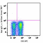
-
APC/Cyanine7 anti-mouse TNF-α
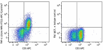
PMA + Ionomycin-stimulated C57BL/6 mouse splenocytes (six-ho... -
PE/Dazzle™ 594 anti-mouse TNF-α
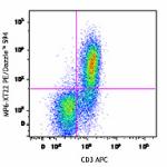
PMA + Ionomycin-stimulated C57BL/6 mouse splenocytes (6-hour... 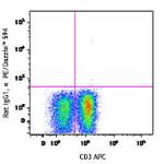
-
Brilliant Violet 711™ anti-mouse TNF-α
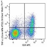
PMA+ Ionomycin-stimulated (six hours) C57BL/6 mouse splenocy... 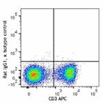
-
Brilliant Violet 750™ anti-mouse TNF-α
PMA + Ionomycin-stimulated C57BL/6 mouse splenocytes (in the... -
GoInVivo™ Purified anti-mouse TNF-α
-
Spark NIR™ 685 anti-mouse TNF-α
Cell Activation Cocktail (with Brefeldin A) stimulated C57BL... -
Biotin anti-mouse TNF-α
-
TotalSeq™-B1529 anti-mouse TNF-α Antibody
-
Alexa Fluor® 488 anti-mouse TNF-α
PMA + Ionomycin-stimulated C57BL/6 mouse splenocytes (in the... 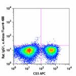
-
Alexa Fluor® 647 anti-mouse TNF-α
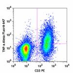
PMA + Ionomycin-stimulated C57BL/6 mouse splenocytes (in the... 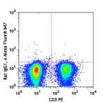
-
TotalSeq™-C1529 anti-mouse TNF-α Antibody
-
Pacific Blue™ anti-mouse TNF-α
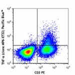
PMA + Ionomycin-stimulated C57BL/6 mouse splenocytes (in the... 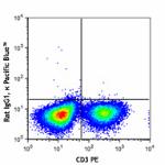
-
PerCP/Cyanine5.5 anti-mouse TNF-α
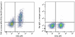
PMA + Ionomycin-stimulated C57BL/6 mouse splenocytes (in the... -
PE/Cyanine7 anti-mouse TNF-α
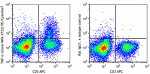
PMA + Ionomycin-stimulated C57BL/6 mouse splenocytes (in the... -
Brilliant Violet 421™ anti-mouse TNF-α
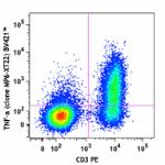
PMA + Ionomycin-stimulated C57BL/6 mouse splenocytes (in the... 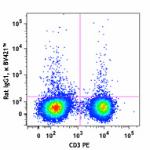
-
Brilliant Violet 605™ anti-mouse TNF-α
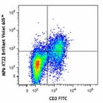
PMA+Ionomycin-stimulated (6 hours) C57BL/6 mouse splenocytes... -
Ultra-LEAF™ Purified anti-mouse TNF-α
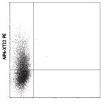
PMA/Ionomycin-stimulated BALB/c T cells were stained with MP... 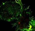
Immortalized murine bone marrow-derived macrophages stimulat... -
Brilliant Violet 650™ anti-mouse TNF-α
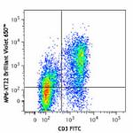
PMA+ Ionomycin-stimulated (6 hours) C57BL/6 mouse splenocyte... -
Alexa Fluor® 700 anti-mouse TNF-α
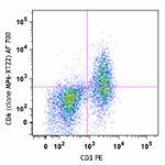
PMA + ionomycin-stimulated (6 hours) C57BL/6 mouse splenocyt... 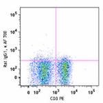
-
Purified anti-mouse TNF-α (Maxpar® Ready)
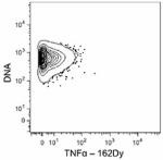
C57BL/6 mouse splenocytes were incubated for 6 hours in medi... 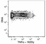
-
Brilliant Violet 510™ anti-mouse TNF-α
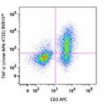
PMA + Ionomycin-stimulated C57BL/6 mouse splenocytes (in the... 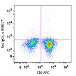
-
APC anti-mouse TNF-α
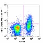
PMA + Ionomycin-stimulated C57BL/6 mouse splenocytes (in the... 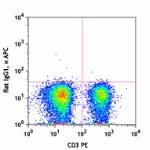
-
FITC anti-mouse TNF-α
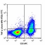
PMA + Ionomycin-stimulated C57BL/6 mouse splenocytes (in the... 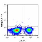
-
PE anti-mouse TNF-α
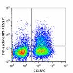
PMA + Ionomycin-stimulated C57BL/6 mouse splenocytes (in the... 
-
Purified anti-mouse TNF-α
PMA/Ionomycin-stimulated BALB/c T cells were stained with MP...
Immortalized murine bone marrow-derived macrophages stimulat...


Follow Us