Anti-β-Amyloid, 1-16 Antibody (Previously Covance catalog# SIG-39300) Anti-β-Amyloid, 1-16 Antibody (Previously Covance catalog# SIG-39300)

Pricing & Availability
Clone
6E10 (See other available formats)
Regulatory Status
RUO
Other Names
AAA, ABETA, ABPP, AD1, APPI, CTFgamma, CVAP, PN-II, PN2, Amyloid beta A4 protein, preA4, protease, peptidase nexin-II, beta-amyloid peptide, alzheimer disease amyloid protein, cerebral vascular amyloid peptide, APP, Amyloid Precursor Protein
Previously
Signet Catalog# 9300-02
Signet Catalog# 9300-05
Signet Catalog# 9300-10
Covance Catalog# SIG-39300
Isotype
Mouse IgG1, κ
Ave. Rating
Submit a Review
Product Citations
publications
A_6E10_ASCITES_betaAmyloid_1-16_WB_050818
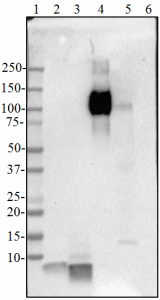
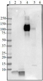
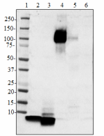
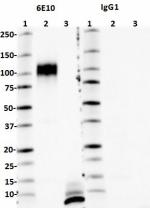
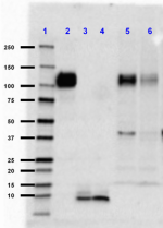
Western blot of anti-β-amyloid, 1-16 antibody (clone 6E10). Lane 1: Molecular weight marker; Lane 2: 50 ng of the human Aβ1-40 peptide; Lane 3: 50 ng of the Aβ1-42 peptide; Lane 4: 50 ng of the recombinant human APP751 protein; Lane 5: 20 µg of the human brain lysate; Lane 6: 50 ng of the rodent Aβ1-42 peptide. The blot was incubated with 1:10,000 dilution of the primary antibody overnight at 4°C, followed by incubation with the HRP-labeled goat anti-mouse IgG (Cat. No. 405306). Enhanced chemiluminescence was used as the detection system.
  • A_6E10_ASCITES_betaAmyloid_1-16_WB_050818
    Western blot of anti-β-amyloid, 1-16 antibody (clone 6E10). Lane 1: Molecular weight marker; Lane 2: 50 ng of the human Aβ1-40 peptide; Lane 3: 50 ng of the Aβ1-42 peptide; Lane 4: 50 ng of the recombinant human APP751 protein; Lane 5: 20 µg of the human brain lysate; Lane 6: 50 ng of the rodent Aβ1-42 peptide. The blot was incubated with 1:10,000 dilution of the primary antibody overnight at 4°C, followed by incubation with the HRP-labeled goat anti-mouse IgG (Cat. No. 405306). Enhanced chemiluminescence was used as the detection system.
  • B_6E10_ASCITES_betaAmyloid_1-16_DirectELISA_050818
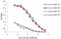
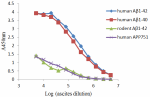
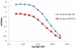
    Direct ELISA of anti-β-amyloid, 1-16 (clone 6E10) antibody binding to the plate-immobilized human Aβ1-40, human Aβ1-42, rodent Aβ1-42 peptides, and recombinant human APP751 protein. ELISA was performed by coating the wells with 100 ng of peptide or recombinant protein. The wells were then incubated with the primary antibody at 37°C for 45 minutes, followed by incubation with HRP labeled goat anti-mouse IgG secondary antibody. TMB (3, 3', 5, 5' tetramethylbenzidine, Cat. No. 421501) was used as the detection system.
  • C_6E10_ASCITES_betaAmyloid_1-16_IHCP_050818
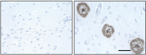
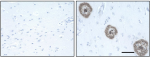
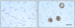
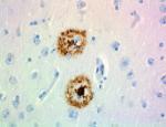
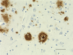
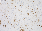
    IHC staining of anti-β-Amyloid, 1-16 antibody (clone 6E10) on formalin-fixed paraffin-embedded normal human (left panel) and Alzheimer’s disease (right panel)brain tissues. Following antigen retrieval using 70% formic acid for 20 minutes, the tissues were incubated with 1:5,000 dilution of the primary antibody for 1 hour at room temperature. BioLegend’s Ultra-Streptavidin (USA) HRP kit (Multi-Species, DAB, Cat. No. 929901) was used for detection followed by hematoxylin counterstaining and bluing solution counterstaining, according to the protocol provided. The images were captured with a 40X objective. Scale bar: 50 µm
Compare all formats
Cat # Size Price Quantity Check Availability Save
803016 500 µL NOK17680
Check Availability


Need larger quantities of this item?
Request Bulk Quote
803017 1 mL NOK29643
Check Availability


Need larger quantities of this item?
Request Bulk Quote
803015 200 µL NOK8696
Check Availability


Need larger quantities of this item?
Request Bulk Quote
803014 50 µL NOK2475
Check Availability


Need larger quantities of this item?
Request Bulk Quote
Description

Alzheimer's disease is characterized by the accumulation of aggregated Aβ peptides in senile plaques and vascular deposits. Aβ peptides are derived from amyloid precursor proteins (APP) through sequential proteolytic cleavage of APP by β-secretases and γ-secretases generating diverse Aβ species. Aβ can aggregate to form soluble oligomeric species and insoluble fibrillar or amorphous assemblies. Some forms of the aggregated peptides are toxic to neurons.

Product Details
Technical Data Sheet (pdf)

Product Details

Verified Reactivity
Human
Antibody Type
Monoclonal
Host Species
Mouse
Preparation
Ascites
Concentration
The concentration is not quantified as this product is sold as undiluted crude mouse ascites fluid. The concentration might vary from lot-to-lot and an estimated concentration would be 1-3 mg/ml.
Storage & Handling
Store at -20°C or below. Upon initial thawing, apportion into working aliquots and store at -20°C or below. Avoid repeated freeze-thaw cycles to prevent denaturing the antibody. Do not store in frost-free freezers.
Application

WB - Quality tested
Direct ELISA, IHC-P - Verified
IHC-F, EM - Reported in the literature, not verified in house

Recommended Usage

Each lot of this antibody is quality control tested by Western blotting. For Western blotting, the suggested dilution of this reagent is 1:1,000 to 1:20,000. For Direct ELISA, the suggested dilution of this reagent is 1:10,000 to 1:100,000. For immunohistochemistry on formalin-fixed paraffin-embedded tissue sections, the suggested dilution of this reagent is 1:1,000 to 1:20,000. It is recommended that the reagent be titrated for optimal performance for each application.

Application Notes

This antibody is reactive to amino acid residue 1-16 of beta amyloid. The epitope lies within amino acids 3-8 of beta amyloid (EFRHDS).

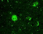
This antibody clone has been reported for use in immunohistochemistry of free-floating sections2,13.

Application References
  1. Thakker DR, et al. 2009. Proc. Natl. Acad. Sci. USA. 106(11):4501-6. (IHC) PubMed
  2. Oddo S, et al. 2005. Proc. Natl. Acad. Sci. USA. 102(8):3046-51. (IHC-other) PubMed
  3. Herzig M, et al. 2004. Nat. Neuro. 7(9):954-959. (WB) PubMed
  4. Zheng Y, et al. 2012. PLoS One 6:39035. (IHC-F) PubMed
  5. Abramowksi D, et al. J Neurosci. 32:1273. (WB) PubMed
  6. Forny-Germano L, et al. 2014. J. Neurosci. 34:13629. (WB, IHC) PubMed
  7. Gowert NS, et al. 2014. PLoS One 2:e90523. (ICC, EM) PubMed
  8. Sandoval-Hernández A, et al. 2015. PLoS One. 10: 0145467. (IHC-F)
  9. Kumar R, et al. 2016. Brain. 139:174-92 (WB)
  10. Miyamoto T, et al. 2016. J. Biol. Chem. 291:1719-34. (WB)
  11. Saito S, et al. 2017. Acta Neuropathol. Commun. 5:26-9. (IHC-P) PubMed
  12. Omata Y, et al. 2016. Aging (Albany NY) 8(3):427. (IHC-P) PubMed
  13. Peng W, et al. 2016. Neurobiol. Dis. 93:215. (IHC-other) PubMed
  14. Mandler M, et al. 2015. PLoS One. e0115237. (WB, IHC, ELISA) PubMed
Product Citations
  1. Fu W, et al. 2018. Exp Ther Med. 128:2252. PubMed
  2. Lana E, et al. 2019. Front Mol Neurosci. 12:176. PubMed
  3. Ren Z, et al. 2020. Mol Med Rep. 22:201. PubMed
  4. Tanaka H, et al. 2020. Nat Commun. 11:507. PubMed
  5. Bishay J, et al. 2022. Sci Rep. 12:15287. PubMed
  6. Li S, et al. 2022. Neural Plast. 2022:3995227. PubMed
  7. Jin J, et al. 2023. Gut Microbes. 15:2167172. PubMed
  8. Yao SY, et al. 2023. Br J Pharmacol. 180:1878. PubMed
  9. Na D, et al. 2023. Front Neurosci. 17:1106570. PubMed
  10. Ren X, et al. 2022. J Neuroinflammation. 19:61. PubMed
  11. Tang T, et al. 2022. J Virol. 96:e0168821. PubMed
  12. Huang S, et al. 2022. FEBS Lett. 596:1059. PubMed
  13. Januário YC, et al. 2022. J Biol Chem. 298:102172. PubMed
  14. Cai C, et al. 2022. J Neurosci. 42:6090. PubMed
  15. Lucero EM, et al. 2022. iScience. 25:105288. PubMed
  16. Hu Y, et al. 2022. Nat Metab. 4:1756. PubMed
  17. Reddy TT, et al. 2023. Molecules. 28: . PubMed
  18. Bathini P, et al. 2023. Brain Behav Immun Health. 28:100568. PubMed
  19. Lin W, et al. 2023. J Neuroinflammation. 20:21. PubMed
  20. Chen SH, et al. 2022. Neurosci Bull. 38:290. PubMed
  21. Arber C, et al. 2019. Brain Commun. 1:fcz024. PubMed
  22. Lai RH, et al. 2021. Aging Cell. 20:e13509. PubMed
  23. NULL, et al. 2021. Mol Imaging Biol. 23:991. PubMed
  24. Cone AS, et al. 2021. Theranostics. 11:8129. PubMed
  25. Forny-Germano L, et al. 2014. J Neurosci. 34:13629-13643. PubMed
  26. Beraldo F, et al. 2016. J Biol Chem. 291: 21945 - 21955. PubMed
  27. Robert J et al. 2017. eLife. 6 pii: e29595. PubMed
  28. Mukherjee J, et al. 2021. Synapse. 75:e22183. PubMed
  29. Zhang H, et al. 2016. J Neurosci. 36: 11837 - 11850. PubMed
  30. Shen Y, et al. 2020. Theranostics. 10:11794. PubMed
  31. Na H, et al. 2021. Am J Alzheimers Dis Other Demen. 36:15333175211012867. PubMed
  32. Shen Q, et al. 2022. Aging Cell. 21:e13573. PubMed
  33. Zheng Y, et al. 2012. PLoS One. 7:e39035. PubMed
  34. Soto-Mercado V, et al. 2020. J Alzheimers Dis. . PubMed
  35. Fujita K, et al. 2016. Sci Rep. 6:31895. PubMed
  36. Suh J, et al. 2020. Cell. 178(5):1159-1175.e17.. PubMed
  37. Nguyen GAH, et al. 2022. Molecules. 27:. PubMed
  38. Shang Y, et al. 2022. Acta Neuropathol. 144:911. PubMed
  39. Gowert N, et al. 2014. PLoS One. 9:e90523. PubMed
  40. Cheng WH, et al. 2019. Alzheimers Res Ther. 11:6. PubMed
  41. Soto-Mercado V, et al. 2021. Biomolecules. 11:. PubMed
  42. Lyra E Silva NM, et al. 2021. Transl Psychiatry. 251:11. PubMed
  43. Benthem SD, et al. 2020. Current Biology. 30(13):2588-2601.e5. PubMed
  44. Zhang J, et al. 2021. Aging Cell. 20:e13380. PubMed
  45. Moelgg K, et al. 2021. Biomolecules. 11:. PubMed
  46. Abramowski D, et al. 2012. J Neurosci. 32:1273-1283. PubMed
  47. Zhang X, et al. 2020. Sci Adv. 6:00. PubMed
  48. Kaur H, et al. 2021. Bioorg Med Chem Lett. 46:128164. PubMed
  49. Qiu Y, et al. 2022. Front Aging Neurosci. 14:896522. PubMed
  50. Kumar S, et al. 2022. NPJ Genom Med. 7:47. PubMed
  51. Casali BT, et al. 2020. Neurobiol Dis. 142:104956. PubMed
  52. Quartey MO, et al. 2021. Sci Rep. 0.757638889. PubMed
  53. Klohs J, et al. 2016. J Cereb Blood Flow Metab. 36: 1614 - 1624. PubMed
  54. Lee CYD et al. 2018. Neuron. 97(5):1032-1048 . PubMed
  55. Ippati S, et al. 2021. Proc Natl Acad Sci U S A. 118:. PubMed
  56. Chan E, et al. 2016. Sci Rep. 6: 26119. PubMed
  57. Boeddrich A et al. 2018. Cell chemical biology. 26(1):109-120 . PubMed
  58. Zhang Y, et al. 2022. Bioact Mater. 11:192. PubMed
  59. Seino S, et al. 2018. Biomed Res. 39:105. PubMed
  60. Lin H, et al. 2022. Front Aging Neurosci. 14:885145. PubMed
  61. Meilandt WJ, et al. 2020. J Neurosci. 40:1956. PubMed
  62. Casali BT, et al. 2018. J Neuroinflammation. 15:43. PubMed
  63. Scholtzova H, et al. 2017. J Neurosci. 37(4):936-959. PubMed
  64. Heiss J, et al. 2016. Cereb Cortex. 10.1093/cercor/bhw188. PubMed
  65. Jang J, et al. 2017. Biochem Biophys Res Commun. 10.1016/j.bbrc.2017.08.127. PubMed
  66. Xiao Zhang et al. 2017. The Journal of Neuroscience. 37(19):4928-4941 . PubMed
  67. Actor-Engel HS, et al. 2021. Eneuro. Online ahead of print. PubMed
  68. Willuweit A, et al. 2021. Front Neurosci. 15:699926. PubMed
  69. Arber C, et al. 2021. Cell Reports. 34(2):108615. PubMed
  70. Song JX, et al. 2020. Aging Cell. 19:e13069. PubMed
  71. Li N, et al. 2019. Neural Regen Res. 14:1037. PubMed
  72. Magalhães J, et al. 2021. Mol Psychiatry. Online ahead of print. PubMed
  73. Dai Y, et al. 2020. Front Cell Dev Biol. 8:593659. PubMed
  74. Porter KN, et al. 2020. Front Aging Neurosci. 12:92. PubMed
  75. Ye T, et al. 2021. Commun Biol. 4:195. PubMed
  76. Ge W, et al. 2021. Am J Transl Res. 13:1471. PubMed
  77. Arber C, et al. 2020. Mol Psychiatry. 25:2919. PubMed
  78. Su Q, et al. 2021. Alzheimers Res Ther. 13:07. PubMed
  79. Soto-Mercado V, et al. 2020. PLoS One. 15:e0221669. PubMed
  80. Kordower JH, et al. 2017. Ann Neurol. 81:46. PubMed
  81. Williamson RL, et al. 2017. J Biol Chem. 292:19873. PubMed
  82. Lai RH, et al. 2022. Aging Cell. 21:e13670. PubMed
  83. Peng D, et al. 2022. Theranostics. 12:3862. PubMed
  84. Li T, et al. 2016. Nat Commun. 7:12082. PubMed
  85. Fu W, et al. 2018. Exp Ther Med. 128:2252. PubMed
  86. Lana E, et al. 2019. Front Mol Neurosci. 12:176. PubMed
  87. Ren Z, et al. 2020. Mol Med Rep. 22:201. PubMed
  88. Tanaka H, et al. 2020. Nat Commun. 11:507. PubMed
  89. Bishay J, et al. 2022. Sci Rep. 12:15287. PubMed
  90. Li S, et al. 2022. Neural Plast. 2022:3995227. PubMed
  91. Jin J, et al. 2023. Gut Microbes. 15:2167172. PubMed
  92. Yao SY, et al. 2023. Br J Pharmacol. 180:1878. PubMed
  93. Na D, et al. 2023. Front Neurosci. 17:1106570. PubMed
  94. Ren X, et al. 2022. J Neuroinflammation. 19:61. PubMed
  95. Tang T, et al. 2022. J Virol. 96:e0168821. PubMed
  96. Huang S, et al. 2022. FEBS Lett. 596:1059. PubMed
  97. Januário YC, et al. 2022. J Biol Chem. 298:102172. PubMed
  98. Cai C, et al. 2022. J Neurosci. 42:6090. PubMed
  99. Lucero EM, et al. 2022. iScience. 25:105288. PubMed
  100. Hu Y, et al. 2022. Nat Metab. 4:1756. PubMed
  101. Reddy TT, et al. 2023. Molecules. 28: . PubMed
  102. Bathini P, et al. 2023. Brain Behav Immun Health. 28:100568. PubMed
  103. Lin W, et al. 2023. J Neuroinflammation. 20:21. PubMed
  104. Chen SH, et al. 2022. Neurosci Bull. 38:290. PubMed
  105. Arber C, et al. 2019. Brain Commun. 1:fcz024. PubMed
  106. Lai RH, et al. 2021. Aging Cell. 20:e13509. PubMed
  107. NULL, et al. 2021. Mol Imaging Biol. 23:991. PubMed
  108. Cone AS, et al. 2021. Theranostics. 11:8129. PubMed
  109. Forny-Germano L, et al. 2014. J Neurosci. 34:13629-13643. PubMed
  110. Beraldo F, et al. 2016. J Biol Chem. 291: 21945 - 21955. PubMed
  111. Robert J et al. 2017. eLife. 6 pii: e29595. PubMed
  112. Mukherjee J, et al. 2021. Synapse. 75:e22183. PubMed
  113. Zhang H, et al. 2016. J Neurosci. 36: 11837 - 11850. PubMed
  114. Shen Y, et al. 2020. Theranostics. 10:11794. PubMed
  115. Na H, et al. 2021. Am J Alzheimers Dis Other Demen. 36:15333175211012867. PubMed
  116. Shen Q, et al. 2022. Aging Cell. 21:e13573. PubMed
  117. Zheng Y, et al. 2012. PLoS One. 7:e39035. PubMed
  118. Soto-Mercado V, et al. 2020. J Alzheimers Dis. . PubMed
  119. Fujita K, et al. 2016. Sci Rep. 6:31895. PubMed
  120. Suh J, et al. 2020. Cell. 178(5):1159-1175.e17.. PubMed
  121. Nguyen GAH, et al. 2022. Molecules. 27:. PubMed
  122. Shang Y, et al. 2022. Acta Neuropathol. 144:911. PubMed
  123. Gowert N, et al. 2014. PLoS One. 9:e90523. PubMed
  124. Cheng WH, et al. 2019. Alzheimers Res Ther. 11:6. PubMed
  125. Soto-Mercado V, et al. 2021. Biomolecules. 11:. PubMed
  126. Lyra E Silva NM, et al. 2021. Transl Psychiatry. 251:11. PubMed
  127. Benthem SD, et al. 2020. Current Biology. 30(13):2588-2601.e5. PubMed
  128. Zhang J, et al. 2021. Aging Cell. 20:e13380. PubMed
  129. Moelgg K, et al. 2021. Biomolecules. 11:. PubMed
  130. Abramowski D, et al. 2012. J Neurosci. 32:1273-1283. PubMed
  131. Zhang X, et al. 2020. Sci Adv. 6:00. PubMed
  132. Kaur H, et al. 2021. Bioorg Med Chem Lett. 46:128164. PubMed
  133. Qiu Y, et al. 2022. Front Aging Neurosci. 14:896522. PubMed
  134. Kumar S, et al. 2022. NPJ Genom Med. 7:47. PubMed
  135. Casali BT, et al. 2020. Neurobiol Dis. 142:104956. PubMed
  136. Quartey MO, et al. 2021. Sci Rep. 0.757638889. PubMed
  137. Klohs J, et al. 2016. J Cereb Blood Flow Metab. 36: 1614 - 1624. PubMed
  138. Lee CYD et al. 2018. Neuron. 97(5):1032-1048 . PubMed
  139. Ippati S, et al. 2021. Proc Natl Acad Sci U S A. 118:. PubMed
  140. Chan E, et al. 2016. Sci Rep. 6: 26119. PubMed
  141. Boeddrich A et al. 2018. Cell chemical biology. 26(1):109-120 . PubMed
  142. Zhang Y, et al. 2022. Bioact Mater. 11:192. PubMed
  143. Seino S, et al. 2018. Biomed Res. 39:105. PubMed
  144. Lin H, et al. 2022. Front Aging Neurosci. 14:885145. PubMed
  145. Meilandt WJ, et al. 2020. J Neurosci. 40:1956. PubMed
  146. Casali BT, et al. 2018. J Neuroinflammation. 15:43. PubMed
  147. Scholtzova H, et al. 2017. J Neurosci. 37(4):936-959. PubMed
  148. Heiss J, et al. 2016. Cereb Cortex. 10.1093/cercor/bhw188. PubMed
  149. Jang J, et al. 2017. Biochem Biophys Res Commun. 10.1016/j.bbrc.2017.08.127. PubMed
  150. Xiao Zhang et al. 2017. The Journal of Neuroscience. 37(19):4928-4941 . PubMed
  151. Actor-Engel HS, et al. 2021. Eneuro. Online ahead of print. PubMed
  152. Willuweit A, et al. 2021. Front Neurosci. 15:699926. PubMed
  153. Arber C, et al. 2021. Cell Reports. 34(2):108615. PubMed
  154. Song JX, et al. 2020. Aging Cell. 19:e13069. PubMed
  155. Li N, et al. 2019. Neural Regen Res. 14:1037. PubMed
  156. Magalhães J, et al. 2021. Mol Psychiatry. Online ahead of print. PubMed
  157. Dai Y, et al. 2020. Front Cell Dev Biol. 8:593659. PubMed
  158. Porter KN, et al. 2020. Front Aging Neurosci. 12:92. PubMed
  159. Ye T, et al. 2021. Commun Biol. 4:195. PubMed
  160. Ge W, et al. 2021. Am J Transl Res. 13:1471. PubMed
  161. Arber C, et al. 2020. Mol Psychiatry. 25:2919. PubMed
  162. Su Q, et al. 2021. Alzheimers Res Ther. 13:07. PubMed
  163. Soto-Mercado V, et al. 2020. PLoS One. 15:e0221669. PubMed
  164. Kordower JH, et al. 2017. Ann Neurol. 81:46. PubMed
  165. Williamson RL, et al. 2017. J Biol Chem. 292:19873. PubMed
  166. Lai RH, et al. 2022. Aging Cell. 21:e13670. PubMed
  167. Peng D, et al. 2022. Theranostics. 12:3862. PubMed
  168. Li T, et al. 2016. Nat Commun. 7:12082. PubMed
RRID
AB_2565329 (BioLegend Cat. No. 803016)
AB_2565327 (BioLegend Cat. No. 803017)
AB_2565328 (BioLegend Cat. No. 803015)
AB_2728527 (BioLegend Cat. No. 803014)

Antigen Details

Structure
Amyloid precursor protein is a 770 amino acid protein with a molecular mass of ~100 kD. According to the UniProtKB database, APP (ID# P05067) has 11 isoforms (34 to ~90 kD) and the 770 form has been designated as the canonical form. Isoform APP695 is the predominant form expressed in neuronal tissue. Isoforms APP751 and APP770 are widely expressed in non-neuronal cells. Isoform APP751 is the most abundant form in T-lymphocytes. Aβ denotes peptides of 36-43 amino acids generated from cleavage of APP by secretases. Aβ has an apparent molecular mass of about 4 kD.
Distribution

Tissue distribution: Primarily nervous system, but also adipose tissue, intestine, muscle.
Cellular distribution: Cytosol, endosomes, nucleus, plasma membrane, extracellular, and golgi apparatus.

Function
The normal function of Aβ is not well understood. Several potential physiological roles have been proposed, including: activation of kinase enzymes; protection against oxidative stress; regulation of cholesterol transport; transcription factor, and as an anti-microbial agent.
Biology Area
Cell Biology, Neurodegeneration, Neuroscience, Protein Misfolding and Aggregation
Molecular Family
APP/β-Amyloid
Antigen References
  1. Kumar A, et al. 2015. Pharmacol. Rep. 67(2):195.
  2. Sadigh-Eteghad S, et al. 2015. Med. Princ. Pract. 24(1):1
  3. Hampel H, et al. 2015. Expert Rev. Neurother. 15(1):83.
  4. Puig KL, et al. 2012.  Exp. Gerontol. 48(7): 608.
  5. Selkoe DJ, et al. 2016. EMBO Mol. Med. 8(6):595.
  6. Walsh DM, et al.  2007. J. Neurochem. 101(5):1172.
Gene ID
351 View all products for this Gene ID
UniProt
View information about beta-Amyloid 1-16 on UniProt.org

Related FAQs

There are no FAQs for this product.
Go To Top Version: 4    Revision Date: 03.12.2021

For Research Use Only. Not for diagnostic or therapeutic use.


This product is supplied subject to the terms and conditions, including the limited license, located at www.biolegend.com/terms) ("Terms") and may be used only as provided in the Terms. Without limiting the foregoing, BioLegend products may not be used for any Commercial Purpose as defined in the Terms, resold in any form, used in manufacturing, or reverse engineered, sequenced, or otherwise studied or used to learn its design or composition without express written approval of BioLegend. Regardless of the information given in this document, user is solely responsible for determining any license requirements necessary for user’s intended use and assumes all risk and liability arising from use of the product. BioLegend is not responsible for patent infringement or any other risks or liabilities whatsoever resulting from the use of its products.


BioLegend, the BioLegend logo, and all other trademarks are property of BioLegend, Inc. or their respective owners, and all rights are reserved.


8999 BioLegend Way, San Diego, CA 92121 www.biolegend.com
Toll-Free Phone: 1-877-Bio-Legend (246-5343) Phone: (858) 768-5800 Fax: (877) 455-9587

This data display is provided for general comparisons between formats.
Your actual data may vary due to variations in samples, target cells, instruments and their settings, staining conditions, and other factors.
If you need assistance with selecting the best format contact our expert technical support team.

Pricing & Availability
Clone
6E10 (See other available formats)
Regulatory Status
RUO
Other Names
AAA, ABETA, ABPP, AD1, APPI, CTFgamma, CVAP, PN-II, PN2, Amyloid beta A4 protein, preA4, protease, peptidase nexin-II, beta-amyloid peptide, alzheimer disease amyloid protein, cerebral vascular amyloid peptide, APP, Amyloid Precursor Protein
Previously
Signet Catalog# 9300-02
Signet Catalog# 9300-05
Signet Catalog# 9300-10
Covance Catalog# SIG-39300
Isotype
Mouse IgG1, κ
Ave. Rating
Submit a Review
Product Citations
publications
A_6E10_ASCITES_betaAmyloid_1-16_WB_050818
Western blot of anti-β-amyloid, 1-16 antibody (clone 6E10). Lane 1: Molecular weight marker; Lane 2: 50 ng of the human Aβ1-40 peptide; Lane 3: 50 ng of the Aβ1-42 peptide; Lane 4: 50 ng of the recombinant human APP751 protein; Lane 5: 20 µg of the human brain lysate; Lane 6: 50 ng of the rodent Aβ1-42 peptide. The blot was incubated with 1:10,000 dilution of the primary antibody overnight at 4°C, followed by incubation with the HRP-labeled goat anti-mouse IgG (Cat. No. 405306). Enhanced chemiluminescence was used as the detection system.
  • A_6E10_ASCITES_betaAmyloid_1-16_WB_050818
    Western blot of anti-β-amyloid, 1-16 antibody (clone 6E10). Lane 1: Molecular weight marker; Lane 2: 50 ng of the human Aβ1-40 peptide; Lane 3: 50 ng of the Aβ1-42 peptide; Lane 4: 50 ng of the recombinant human APP751 protein; Lane 5: 20 µg of the human brain lysate; Lane 6: 50 ng of the rodent Aβ1-42 peptide. The blot was incubated with 1:10,000 dilution of the primary antibody overnight at 4°C, followed by incubation with the HRP-labeled goat anti-mouse IgG (Cat. No. 405306). Enhanced chemiluminescence was used as the detection system.
  • B_6E10_ASCITES_betaAmyloid_1-16_DirectELISA_050818
    Direct ELISA of anti-β-amyloid, 1-16 (clone 6E10) antibody binding to the plate-immobilized human Aβ1-40, human Aβ1-42, rodent Aβ1-42 peptides, and recombinant human APP751 protein. ELISA was performed by coating the wells with 100 ng of peptide or recombinant protein. The wells were then incubated with the primary antibody at 37°C for 45 minutes, followed by incubation with HRP labeled goat anti-mouse IgG secondary antibody. TMB (3, 3', 5, 5' tetramethylbenzidine, Cat. No. 421501) was used as the detection system.
  • C_6E10_ASCITES_betaAmyloid_1-16_IHCP_050818
    IHC staining of anti-β-Amyloid, 1-16 antibody (clone 6E10) on formalin-fixed paraffin-embedded normal human (left panel) and Alzheimer’s disease (right panel)brain tissues. Following antigen retrieval using 70% formic acid for 20 minutes, the tissues were incubated with 1:5,000 dilution of the primary antibody for 1 hour at room temperature. BioLegend’s Ultra-Streptavidin (USA) HRP kit (Multi-Species, DAB, Cat. No. 929901) was used for detection followed by hematoxylin counterstaining and bluing solution counterstaining, according to the protocol provided. The images were captured with a 40X objective. Scale bar: 50 µm
Compare all formats
Cat # Size Price Quantity Check Availability Save
803016 500 µL NOK17680
Check Availability


Need larger quantities of this item?
Request Bulk Quote
803017 1 mL NOK29643
Check Availability


Need larger quantities of this item?
Request Bulk Quote
803015 200 µL NOK8696
Check Availability


Need larger quantities of this item?
Request Bulk Quote
803014 50 µL NOK2475
Check Availability


Need larger quantities of this item?
Request Bulk Quote
Description

Alzheimer's disease is characterized by the accumulation of aggregated Aβ peptides in senile plaques and vascular deposits. Aβ peptides are derived from amyloid precursor proteins (APP) through sequential proteolytic cleavage of APP by β-secretases and γ-secretases generating diverse Aβ species. Aβ can aggregate to form soluble oligomeric species and insoluble fibrillar or amorphous assemblies. Some forms of the aggregated peptides are toxic to neurons.

Product Details
Technical Data Sheet (pdf)

Product Details

Verified Reactivity
Human
Antibody Type
Monoclonal
Host Species
Mouse
Preparation
Ascites
Concentration
The concentration is not quantified as this product is sold as undiluted crude mouse ascites fluid. The concentration might vary from lot-to-lot and an estimated concentration would be 1-3 mg/ml.
Storage & Handling
Store at -20°C or below. Upon initial thawing, apportion into working aliquots and store at -20°C or below. Avoid repeated freeze-thaw cycles to prevent denaturing the antibody. Do not store in frost-free freezers.
Application

WB - Quality tested
Direct ELISA, IHC-P - Verified
IHC-F, EM - Reported in the literature, not verified in house

Recommended Usage

Each lot of this antibody is quality control tested by Western blotting. For Western blotting, the suggested dilution of this reagent is 1:1,000 to 1:20,000. For Direct ELISA, the suggested dilution of this reagent is 1:10,000 to 1:100,000. For immunohistochemistry on formalin-fixed paraffin-embedded tissue sections, the suggested dilution of this reagent is 1:1,000 to 1:20,000. It is recommended that the reagent be titrated for optimal performance for each application.

Application Notes

This antibody is reactive to amino acid residue 1-16 of beta amyloid. The epitope lies within amino acids 3-8 of beta amyloid (EFRHDS).

This antibody clone has been reported for use in immunohistochemistry of free-floating sections2,13.

Application References
  1. Thakker DR, et al. 2009. Proc. Natl. Acad. Sci. USA. 106(11):4501-6. (IHC) PubMed
  2. Oddo S, et al. 2005. Proc. Natl. Acad. Sci. USA. 102(8):3046-51. (IHC-other) PubMed
  3. Herzig M, et al. 2004. Nat. Neuro. 7(9):954-959. (WB) PubMed
  4. Zheng Y, et al. 2012. PLoS One 6:39035. (IHC-F) PubMed
  5. Abramowksi D, et al. J Neurosci. 32:1273. (WB) PubMed
  6. Forny-Germano L, et al. 2014. J. Neurosci. 34:13629. (WB, IHC) PubMed
  7. Gowert NS, et al. 2014. PLoS One 2:e90523. (ICC, EM) PubMed
  8. Sandoval-Hernández A, et al. 2015. PLoS One. 10: 0145467. (IHC-F)
  9. Kumar R, et al. 2016. Brain. 139:174-92 (WB)
  10. Miyamoto T, et al. 2016. J. Biol. Chem. 291:1719-34. (WB)
  11. Saito S, et al. 2017. Acta Neuropathol. Commun. 5:26-9. (IHC-P) PubMed
  12. Omata Y, et al. 2016. Aging (Albany NY) 8(3):427. (IHC-P) PubMed
  13. Peng W, et al. 2016. Neurobiol. Dis. 93:215. (IHC-other) PubMed
  14. Mandler M, et al. 2015. PLoS One. e0115237. (WB, IHC, ELISA) PubMed
Product Citations
  1. Fu W, et al. 2018. Exp Ther Med. 128:2252. PubMed
  2. Lana E, et al. 2019. Front Mol Neurosci. 12:176. PubMed
  3. Ren Z, et al. 2020. Mol Med Rep. 22:201. PubMed
  4. Tanaka H, et al. 2020. Nat Commun. 11:507. PubMed
  5. Bishay J, et al. 2022. Sci Rep. 12:15287. PubMed
  6. Li S, et al. 2022. Neural Plast. 2022:3995227. PubMed
  7. Jin J, et al. 2023. Gut Microbes. 15:2167172. PubMed
  8. Yao SY, et al. 2023. Br J Pharmacol. 180:1878. PubMed
  9. Na D, et al. 2023. Front Neurosci. 17:1106570. PubMed
  10. Ren X, et al. 2022. J Neuroinflammation. 19:61. PubMed
  11. Tang T, et al. 2022. J Virol. 96:e0168821. PubMed
  12. Huang S, et al. 2022. FEBS Lett. 596:1059. PubMed
  13. Januário YC, et al. 2022. J Biol Chem. 298:102172. PubMed
  14. Cai C, et al. 2022. J Neurosci. 42:6090. PubMed
  15. Lucero EM, et al. 2022. iScience. 25:105288. PubMed
  16. Hu Y, et al. 2022. Nat Metab. 4:1756. PubMed
  17. Reddy TT, et al. 2023. Molecules. 28: . PubMed
  18. Bathini P, et al. 2023. Brain Behav Immun Health. 28:100568. PubMed
  19. Lin W, et al. 2023. J Neuroinflammation. 20:21. PubMed
  20. Chen SH, et al. 2022. Neurosci Bull. 38:290. PubMed
  21. Arber C, et al. 2019. Brain Commun. 1:fcz024. PubMed
  22. Lai RH, et al. 2021. Aging Cell. 20:e13509. PubMed
  23. NULL, et al. 2021. Mol Imaging Biol. 23:991. PubMed
  24. Cone AS, et al. 2021. Theranostics. 11:8129. PubMed
  25. Forny-Germano L, et al. 2014. J Neurosci. 34:13629-13643. PubMed
  26. Beraldo F, et al. 2016. J Biol Chem. 291: 21945 - 21955. PubMed
  27. Robert J et al. 2017. eLife. 6 pii: e29595. PubMed
  28. Mukherjee J, et al. 2021. Synapse. 75:e22183. PubMed
  29. Zhang H, et al. 2016. J Neurosci. 36: 11837 - 11850. PubMed
  30. Shen Y, et al. 2020. Theranostics. 10:11794. PubMed
  31. Na H, et al. 2021. Am J Alzheimers Dis Other Demen. 36:15333175211012867. PubMed
  32. Shen Q, et al. 2022. Aging Cell. 21:e13573. PubMed
  33. Zheng Y, et al. 2012. PLoS One. 7:e39035. PubMed
  34. Soto-Mercado V, et al. 2020. J Alzheimers Dis. . PubMed
  35. Fujita K, et al. 2016. Sci Rep. 6:31895. PubMed
  36. Suh J, et al. 2020. Cell. 178(5):1159-1175.e17.. PubMed
  37. Nguyen GAH, et al. 2022. Molecules. 27:. PubMed
  38. Shang Y, et al. 2022. Acta Neuropathol. 144:911. PubMed
  39. Gowert N, et al. 2014. PLoS One. 9:e90523. PubMed
  40. Cheng WH, et al. 2019. Alzheimers Res Ther. 11:6. PubMed
  41. Soto-Mercado V, et al. 2021. Biomolecules. 11:. PubMed
  42. Lyra E Silva NM, et al. 2021. Transl Psychiatry. 251:11. PubMed
  43. Benthem SD, et al. 2020. Current Biology. 30(13):2588-2601.e5. PubMed
  44. Zhang J, et al. 2021. Aging Cell. 20:e13380. PubMed
  45. Moelgg K, et al. 2021. Biomolecules. 11:. PubMed
  46. Abramowski D, et al. 2012. J Neurosci. 32:1273-1283. PubMed
  47. Zhang X, et al. 2020. Sci Adv. 6:00. PubMed
  48. Kaur H, et al. 2021. Bioorg Med Chem Lett. 46:128164. PubMed
  49. Qiu Y, et al. 2022. Front Aging Neurosci. 14:896522. PubMed
  50. Kumar S, et al. 2022. NPJ Genom Med. 7:47. PubMed
  51. Casali BT, et al. 2020. Neurobiol Dis. 142:104956. PubMed
  52. Quartey MO, et al. 2021. Sci Rep. 0.757638889. PubMed
  53. Klohs J, et al. 2016. J Cereb Blood Flow Metab. 36: 1614 - 1624. PubMed
  54. Lee CYD et al. 2018. Neuron. 97(5):1032-1048 . PubMed
  55. Ippati S, et al. 2021. Proc Natl Acad Sci U S A. 118:. PubMed
  56. Chan E, et al. 2016. Sci Rep. 6: 26119. PubMed
  57. Boeddrich A et al. 2018. Cell chemical biology. 26(1):109-120 . PubMed
  58. Zhang Y, et al. 2022. Bioact Mater. 11:192. PubMed
  59. Seino S, et al. 2018. Biomed Res. 39:105. PubMed
  60. Lin H, et al. 2022. Front Aging Neurosci. 14:885145. PubMed
  61. Meilandt WJ, et al. 2020. J Neurosci. 40:1956. PubMed
  62. Casali BT, et al. 2018. J Neuroinflammation. 15:43. PubMed
  63. Scholtzova H, et al. 2017. J Neurosci. 37(4):936-959. PubMed
  64. Heiss J, et al. 2016. Cereb Cortex. 10.1093/cercor/bhw188. PubMed
  65. Jang J, et al. 2017. Biochem Biophys Res Commun. 10.1016/j.bbrc.2017.08.127. PubMed
  66. Xiao Zhang et al. 2017. The Journal of Neuroscience. 37(19):4928-4941 . PubMed
  67. Actor-Engel HS, et al. 2021. Eneuro. Online ahead of print. PubMed
  68. Willuweit A, et al. 2021. Front Neurosci. 15:699926. PubMed
  69. Arber C, et al. 2021. Cell Reports. 34(2):108615. PubMed
  70. Song JX, et al. 2020. Aging Cell. 19:e13069. PubMed
  71. Li N, et al. 2019. Neural Regen Res. 14:1037. PubMed
  72. Magalhães J, et al. 2021. Mol Psychiatry. Online ahead of print. PubMed
  73. Dai Y, et al. 2020. Front Cell Dev Biol. 8:593659. PubMed
  74. Porter KN, et al. 2020. Front Aging Neurosci. 12:92. PubMed
  75. Ye T, et al. 2021. Commun Biol. 4:195. PubMed
  76. Ge W, et al. 2021. Am J Transl Res. 13:1471. PubMed
  77. Arber C, et al. 2020. Mol Psychiatry. 25:2919. PubMed
  78. Su Q, et al. 2021. Alzheimers Res Ther. 13:07. PubMed
  79. Soto-Mercado V, et al. 2020. PLoS One. 15:e0221669. PubMed
  80. Kordower JH, et al. 2017. Ann Neurol. 81:46. PubMed
  81. Williamson RL, et al. 2017. J Biol Chem. 292:19873. PubMed
  82. Lai RH, et al. 2022. Aging Cell. 21:e13670. PubMed
  83. Peng D, et al. 2022. Theranostics. 12:3862. PubMed
  84. Li T, et al. 2016. Nat Commun. 7:12082. PubMed
  85. Fu W, et al. 2018. Exp Ther Med. 128:2252. PubMed
  86. Lana E, et al. 2019. Front Mol Neurosci. 12:176. PubMed
  87. Ren Z, et al. 2020. Mol Med Rep. 22:201. PubMed
  88. Tanaka H, et al. 2020. Nat Commun. 11:507. PubMed
  89. Bishay J, et al. 2022. Sci Rep. 12:15287. PubMed
  90. Li S, et al. 2022. Neural Plast. 2022:3995227. PubMed
  91. Jin J, et al. 2023. Gut Microbes. 15:2167172. PubMed
  92. Yao SY, et al. 2023. Br J Pharmacol. 180:1878. PubMed
  93. Na D, et al. 2023. Front Neurosci. 17:1106570. PubMed
  94. Ren X, et al. 2022. J Neuroinflammation. 19:61. PubMed
  95. Tang T, et al. 2022. J Virol. 96:e0168821. PubMed
  96. Huang S, et al. 2022. FEBS Lett. 596:1059. PubMed
  97. Januário YC, et al. 2022. J Biol Chem. 298:102172. PubMed
  98. Cai C, et al. 2022. J Neurosci. 42:6090. PubMed
  99. Lucero EM, et al. 2022. iScience. 25:105288. PubMed
  100. Hu Y, et al. 2022. Nat Metab. 4:1756. PubMed
  101. Reddy TT, et al. 2023. Molecules. 28: . PubMed
  102. Bathini P, et al. 2023. Brain Behav Immun Health. 28:100568. PubMed
  103. Lin W, et al. 2023. J Neuroinflammation. 20:21. PubMed
  104. Chen SH, et al. 2022. Neurosci Bull. 38:290. PubMed
  105. Arber C, et al. 2019. Brain Commun. 1:fcz024. PubMed
  106. Lai RH, et al. 2021. Aging Cell. 20:e13509. PubMed
  107. NULL, et al. 2021. Mol Imaging Biol. 23:991. PubMed
  108. Cone AS, et al. 2021. Theranostics. 11:8129. PubMed
  109. Forny-Germano L, et al. 2014. J Neurosci. 34:13629-13643. PubMed
  110. Beraldo F, et al. 2016. J Biol Chem. 291: 21945 - 21955. PubMed
  111. Robert J et al. 2017. eLife. 6 pii: e29595. PubMed
  112. Mukherjee J, et al. 2021. Synapse. 75:e22183. PubMed
  113. Zhang H, et al. 2016. J Neurosci. 36: 11837 - 11850. PubMed
  114. Shen Y, et al. 2020. Theranostics. 10:11794. PubMed
  115. Na H, et al. 2021. Am J Alzheimers Dis Other Demen. 36:15333175211012867. PubMed
  116. Shen Q, et al. 2022. Aging Cell. 21:e13573. PubMed
  117. Zheng Y, et al. 2012. PLoS One. 7:e39035. PubMed
  118. Soto-Mercado V, et al. 2020. J Alzheimers Dis. . PubMed
  119. Fujita K, et al. 2016. Sci Rep. 6:31895. PubMed
  120. Suh J, et al. 2020. Cell. 178(5):1159-1175.e17.. PubMed
  121. Nguyen GAH, et al. 2022. Molecules. 27:. PubMed
  122. Shang Y, et al. 2022. Acta Neuropathol. 144:911. PubMed
  123. Gowert N, et al. 2014. PLoS One. 9:e90523. PubMed
  124. Cheng WH, et al. 2019. Alzheimers Res Ther. 11:6. PubMed
  125. Soto-Mercado V, et al. 2021. Biomolecules. 11:. PubMed
  126. Lyra E Silva NM, et al. 2021. Transl Psychiatry. 251:11. PubMed
  127. Benthem SD, et al. 2020. Current Biology. 30(13):2588-2601.e5. PubMed
  128. Zhang J, et al. 2021. Aging Cell. 20:e13380. PubMed
  129. Moelgg K, et al. 2021. Biomolecules. 11:. PubMed
  130. Abramowski D, et al. 2012. J Neurosci. 32:1273-1283. PubMed
  131. Zhang X, et al. 2020. Sci Adv. 6:00. PubMed
  132. Kaur H, et al. 2021. Bioorg Med Chem Lett. 46:128164. PubMed
  133. Qiu Y, et al. 2022. Front Aging Neurosci. 14:896522. PubMed
  134. Kumar S, et al. 2022. NPJ Genom Med. 7:47. PubMed
  135. Casali BT, et al. 2020. Neurobiol Dis. 142:104956. PubMed
  136. Quartey MO, et al. 2021. Sci Rep. 0.757638889. PubMed
  137. Klohs J, et al. 2016. J Cereb Blood Flow Metab. 36: 1614 - 1624. PubMed
  138. Lee CYD et al. 2018. Neuron. 97(5):1032-1048 . PubMed
  139. Ippati S, et al. 2021. Proc Natl Acad Sci U S A. 118:. PubMed
  140. Chan E, et al. 2016. Sci Rep. 6: 26119. PubMed
  141. Boeddrich A et al. 2018. Cell chemical biology. 26(1):109-120 . PubMed
  142. Zhang Y, et al. 2022. Bioact Mater. 11:192. PubMed
  143. Seino S, et al. 2018. Biomed Res. 39:105. PubMed
  144. Lin H, et al. 2022. Front Aging Neurosci. 14:885145. PubMed
  145. Meilandt WJ, et al. 2020. J Neurosci. 40:1956. PubMed
  146. Casali BT, et al. 2018. J Neuroinflammation. 15:43. PubMed
  147. Scholtzova H, et al. 2017. J Neurosci. 37(4):936-959. PubMed
  148. Heiss J, et al. 2016. Cereb Cortex. 10.1093/cercor/bhw188. PubMed
  149. Jang J, et al. 2017. Biochem Biophys Res Commun. 10.1016/j.bbrc.2017.08.127. PubMed
  150. Xiao Zhang et al. 2017. The Journal of Neuroscience. 37(19):4928-4941 . PubMed
  151. Actor-Engel HS, et al. 2021. Eneuro. Online ahead of print. PubMed
  152. Willuweit A, et al. 2021. Front Neurosci. 15:699926. PubMed
  153. Arber C, et al. 2021. Cell Reports. 34(2):108615. PubMed
  154. Song JX, et al. 2020. Aging Cell. 19:e13069. PubMed
  155. Li N, et al. 2019. Neural Regen Res. 14:1037. PubMed
  156. Magalhães J, et al. 2021. Mol Psychiatry. Online ahead of print. PubMed
  157. Dai Y, et al. 2020. Front Cell Dev Biol. 8:593659. PubMed
  158. Porter KN, et al. 2020. Front Aging Neurosci. 12:92. PubMed
  159. Ye T, et al. 2021. Commun Biol. 4:195. PubMed
  160. Ge W, et al. 2021. Am J Transl Res. 13:1471. PubMed
  161. Arber C, et al. 2020. Mol Psychiatry. 25:2919. PubMed
  162. Su Q, et al. 2021. Alzheimers Res Ther. 13:07. PubMed
  163. Soto-Mercado V, et al. 2020. PLoS One. 15:e0221669. PubMed
  164. Kordower JH, et al. 2017. Ann Neurol. 81:46. PubMed
  165. Williamson RL, et al. 2017. J Biol Chem. 292:19873. PubMed
  166. Lai RH, et al. 2022. Aging Cell. 21:e13670. PubMed
  167. Peng D, et al. 2022. Theranostics. 12:3862. PubMed
  168. Li T, et al. 2016. Nat Commun. 7:12082. PubMed
RRID
AB_2565329 (BioLegend Cat. No. 803016)
AB_2565327 (BioLegend Cat. No. 803017)
AB_2565328 (BioLegend Cat. No. 803015)
AB_2728527 (BioLegend Cat. No. 803014)

Antigen Details

Structure
Amyloid precursor protein is a 770 amino acid protein with a molecular mass of ~100 kD. According to the UniProtKB database, APP (ID# P05067) has 11 isoforms (34 to ~90 kD) and the 770 form has been designated as the canonical form. Isoform APP695 is the predominant form expressed in neuronal tissue. Isoforms APP751 and APP770 are widely expressed in non-neuronal cells. Isoform APP751 is the most abundant form in T-lymphocytes. Aβ denotes peptides of 36-43 amino acids generated from cleavage of APP by secretases. Aβ has an apparent molecular mass of about 4 kD.
Distribution

Tissue distribution: Primarily nervous system, but also adipose tissue, intestine, muscle.
Cellular distribution: Cytosol, endosomes, nucleus, plasma membrane, extracellular, and golgi apparatus.

Function
The normal function of Aβ is not well understood. Several potential physiological roles have been proposed, including: activation of kinase enzymes; protection against oxidative stress; regulation of cholesterol transport; transcription factor, and as an anti-microbial agent.
Biology Area
Cell Biology, Neurodegeneration, Neuroscience, Protein Misfolding and Aggregation
Molecular Family
APP/β-Amyloid
Antigen References
  1. Kumar A, et al. 2015. Pharmacol. Rep. 67(2):195.
  2. Sadigh-Eteghad S, et al. 2015. Med. Princ. Pract. 24(1):1
  3. Hampel H, et al. 2015. Expert Rev. Neurother. 15(1):83.
  4. Puig KL, et al. 2012.  Exp. Gerontol. 48(7): 608.
  5. Selkoe DJ, et al. 2016. EMBO Mol. Med. 8(6):595.
  6. Walsh DM, et al.  2007. J. Neurochem. 101(5):1172.
Gene ID
351 View all products for this Gene ID
UniProt
View information about beta-Amyloid 1-16 on UniProt.org
Go To Top Version: 4    Revision Date: 03.12.2021

For Research Use Only. Not for diagnostic or therapeutic use.


This product is supplied subject to the terms and conditions, including the limited license, located at www.biolegend.com/terms) ("Terms") and may be used only as provided in the Terms. Without limiting the foregoing, BioLegend products may not be used for any Commercial Purpose as defined in the Terms, resold in any form, used in manufacturing, or reverse engineered, sequenced, or otherwise studied or used to learn its design or composition without express written approval of BioLegend. Regardless of the information given in this document, user is solely responsible for determining any license requirements necessary for user’s intended use and assumes all risk and liability arising from use of the product. BioLegend is not responsible for patent infringement or any other risks or liabilities whatsoever resulting from the use of its products.


BioLegend, the BioLegend logo, and all other trademarks are property of BioLegend, Inc. or their respective owners, and all rights are reserved.


8999 BioLegend Way, San Diego, CA 92121 www.biolegend.com
Toll-Free Phone: 1-877-Bio-Legend (246-5343) Phone: (858) 768-5800 Fax: (877) 455-9587

This data display is provided for general comparisons between formats.
Your actual data may vary due to variations in samples, target cells, instruments and their settings, staining conditions, and other factors.
If you need assistance with selecting the best format contact our expert technical support team.

ProductsHere

Login / Register
Remember me
Forgot your password? Reset password?
Create an Account