FITC anti-human CD3 Antibody FITC anti-human CD3 Antibody

Pricing & Availability
Clone
OKT3 (See other available formats)
Regulatory Status
RUO
Workshop
HCDM listed
Other Names
T3, CD3ε
Isotype
Mouse IgG2a, κ
Ave. Rating
Submit a Review
Product Citations
publications
OKT3_FITC_053107
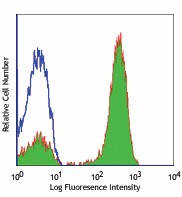
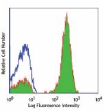
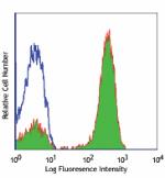
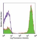
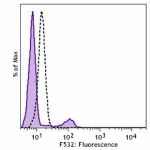
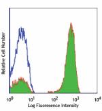
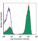
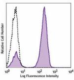
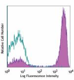
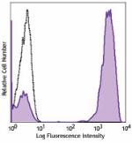
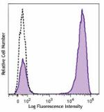
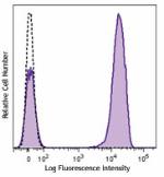
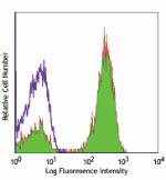
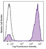
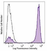
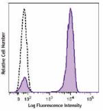
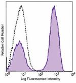
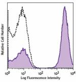
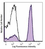
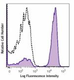
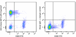
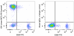
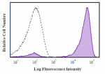
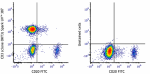
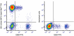
Human peripheral blood lymphocytes stained with OKT3 FITC
  • OKT3_FITC_053107
    Human peripheral blood lymphocytes stained with OKT3 FITC
Compare all formats See FITC spectral data
Cat # Size Price Quantity Check Availability Save
317305 25 tests NOK231
Check Availability


Need larger quantities of this item?
Request Bulk Quote
317306 100 tests NOK454
Check Availability


Need larger quantities of this item?
Request Bulk Quote
Description

CD3ε is a 20 kD chain of the CD3/T cell receptor (TCR) complex, which is composed of two CD3ε, one CD3γ, one CD3δ, one CD3ζ (CD247), and a T cell receptor (α/β or γ/δ) heterodimer. It is found on all mature T lymphocytes, NK T cells, and some thymocytes. CD3, also known as T3, is a member of the immunoglobulin superfamily that plays a role in antigen recognition, signal transduction, and T cell activation.

Product Details
Technical Data Sheet (pdf)

Product Details

Verified Reactivity
Human
Antibody Type
Monoclonal
Host Species
Mouse
Formulation
Phosphate-buffered solution, pH 7.2, containing 0.09% sodium azide and BSA (origin USA)
Preparation
The antibody was purified by affinity chromatography, and conjugated with FITC under optimal conditions.
Concentration
Lot-specific (to obtain lot-specific concentration and expiration, please enter the lot number in our Certificate of Analysis online tool.)
Storage & Handling
The antibody solution should be stored undiluted between 2°C and 8°C, and protected from prolonged exposure to light. Do not freeze.
Application

FC - Quality tested

Recommended Usage

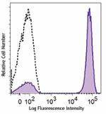
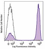
Each lot of this antibody is quality control tested by immunofluorescent staining with flow cytometric analysis. For flow cytometric staining, the suggested use of this reagent is 5 µl per million cells in 100 µl staining volume or 5 µl per 100 µl of whole blood.

Excitation Laser
Blue Laser (488 nm)
Application Notes

The OKT3 monoclonal antibody reacts with an epitope on the epsilon-subunit within the human CD3 complex.

Clone OKT3 can block the binding of clones SK7 and UCHT1.4 The OKT3 antibody is able to induce T cell activation. Additional reported applications (for the relevant formats) include: immunohistochemical staining of acetone-fixed frozen sections and activation of T cells. The LEAF™ purified antibody (Endotoxin <0.1 EU/µg, Azide-Free, 0.2 µm filtered) is recommended for functional assays (Cat. No. 317304). For highly sensitive assays, we recommend Ultra-LEAF™ purified antibody (Cat. No. 317326) with a lower endotoxin limit than standard LEAF™ purified antibodies (Endotoxin <0.01 EU/µg).

Application References
  1. Schlossman S, et al. Eds. 1995. Leucocyte Typing V. Oxford University Press. New York.
  2. Knapp W. 1989. Leucocyte Typing IV. Oxford University Press New York.
  3. Barclay N, et al. 1997. The Leucocyte Antigen Facts Book. Academic Press Inc. San Diego.
  4. Li B, et al. 2005. Immunology 116:487.
  5. Jeong HY, et al. 2008. J. Leuckocyte Biol. 83:755. PubMed
  6. Alter G, et al. 2008. J. Virol. 82:9668. PubMed
  7. Manevich-Mendelson E, et al. 2009. Blood 114:2344. PubMed
  8. Pinto JP, et al. 2010. Immunology. 130:217. PubMed
  9. Biggs MJ, et al. 2011. J. R. Soc. Interface. 8:1462. PubMed
Product Citations
  1. Gkika E, et al. 2023. NPJ Precis Oncol. 7:24. PubMed
  2. Li D, et al. 2023. Int J Oncol. 62:. PubMed
  3. Li L, et al. 2023. Heliyon. 9:e14219. PubMed
  4. Deng G, et al. 2023. Cancers (Basel). 15:. PubMed
  5. Kim JH, et al. 2020. Sci Rep. 10:1835. PubMed
  6. Zhang T, et al. 2022. Am J Reprod Immunol. 87:e13524. PubMed
  7. Tessaro FHG, et al. 2022. Cell Rep. 39:110977. PubMed
  8. Deepak HB, et al. 2022. Indian J Pharmacol. 54:183. PubMed
  9. Rosato F, et al. 2022. J Transl Med. 20:578. PubMed
  10. Zhang W, et al. 2022. Front Cell Dev Biol. 10:842813. PubMed
  11. Zitti B, et al. 2017. Sci Rep. 10.1038/s41598-017-10403-0. PubMed
  12. Afolabi LO, et al. 2021. Front Immunol. 12:701671. PubMed
  13. Guo X, et al. 2016. Mol Ther Methods Clin Dev. 3:15054. PubMed
  14. Want MY, et al. 2019. Oncoimmunology. 8:e1586042. PubMed
  15. Lugo-Reyes SO, et al. 2021. J Clin Immunol. 41:1291. PubMed
  16. Hess NJ, et al. 2020. J Immunol. 205:272. PubMed
  17. Clayton K, et al. 2014. J Immunol. 192:782. PubMed
  18. Catapano M, et al. 2020. J Invest Dermatol. 140:816. PubMed
  19. Shi Z, et al. 2021. Ann Clin Transl Neurol. 8:43. PubMed
  20. Wang Y, et al. 2019. J Transl Med. 17:93. PubMed
  21. Wu X, et al. 2020. Cancers (Basel). 12:. PubMed
  22. Moen IN, et al. 2021. Cell Death Discov. 7:36. PubMed
  23. Yamada E, et al. 2021. Gut Microbes. 13:1997292. PubMed
  24. Zhang B, et al. 2022. J Exp Med. 219:. PubMed
  25. Dai Q, et al. 2020. Front Immunol. 11:539654. PubMed
  26. Lin L, et al. 2018. Oncol Lett. 16:1259. PubMed
  27. Sun Y, et al. 2022. Nat Commun. 13:3916. PubMed
  28. Zhang Z, et al. 2021. Acta Pharm Sin B. 11:1965. PubMed
  29. Celik H, et al. 2021. Cancer Discov. 11:3126. PubMed
  30. Giesecke C, et al. 2017. Cytometry A. 10.1002/cyto.a.23250. PubMed
  31. Fujigaki J, et al. 2015. PLoS One. 10: 0132521. PubMed
  32. Gillissen MA, et al. 2018. Blood. 131:131. PubMed
  33. Kim K, et al. 2019. Nat Microbiol. 1.586111111. PubMed
  34. Sperk M, et al. 2021. iScience. 24:102111. PubMed
  35. Nakasone H, et al. 2021. Commun Biol. 4:1177. PubMed
  36. Wu X, et al. 2021. Front Immunol. 12:731767. PubMed
  37. Jeyanathan M, et al. 2022. JCI Insight. 7:. PubMed
  38. Omer OS, et al. 2020. Methods Mol Biol. 2121:199. PubMed
  39. James KR, et al. 2020. Nat Immunol. 1.113194444. PubMed
  40. Hazenberg MD, et al. 2019. Blood Adv. 2.659722222. PubMed
  41. Chulpanova DS, et al. 2021. Biology (Basel). 10:. PubMed
  42. Golebski K, et al. 2021. Immunity. 54(2):291-307.e7. PubMed
  43. Agashe VV, et al. 2018. J Immunol. 201:772. PubMed
  44. Oyer JL, et al. 2022. Front Immunol. 13:861681. PubMed
  45. Zhang CR, et al. 2022. Blood Cancer Discov. 3:220. PubMed
  46. Cheng J, et al. 2022. iScience. 25:103588. PubMed
  47. Tsai HI, et al. 2021. EMBO Mol Med. 13:e12834. PubMed
  48. Zhang W, et al. 2022. J Transl Med. 20:558. PubMed
  49. Aagaard K, et al. 2017. Sci Rep. 7:41389. PubMed
  50. Miyamae Y, et al. 2020. Cell Chemical Biology. 27(12):1573-1581.e3. PubMed
  51. Winkler C, et al. 2021. JCI Insight. 6:. PubMed
  52. Akusjärvi SS, et al. 2022. iScience. 25:103607. PubMed
  53. Zhou Z, et al. 2022. J Gastrointest Oncol. 13:732. PubMed
  54. Zhao B, et al. 2013. PLoS One. 8:77708. PubMed
  55. Bal S, et al. 2016. Nat Immunol. 10.1038/ni.3444. PubMed
  56. Gopalakrishnapillai A, et al. 2021. Cancers (Basel). 13:. PubMed
  57. Loo Yau H, et al. 2021. STAR Protocols. 2(2):100549. PubMed
  58. Shikhagaie M, et al. 2017. Cell Rep. 18(7):1761-1773. PubMed
  59. Sullivan KD et al. 2016. eLife. 5 pii: e16220. PubMed
  60. Ochi T, et al. 2011. Blood. 118:1495. PubMed
  61. Svensson Akusjärvi S, et al. 2022. Commun Biol. 5:357. PubMed
  62. Fierle JK, et al. 2021. Cell Reports Medicine. 2(8):100362. PubMed
  63. Gkika E, et al. 2023. NPJ Precis Oncol. 7:24. PubMed
  64. Li D, et al. 2023. Int J Oncol. 62:. PubMed
  65. Li L, et al. 2023. Heliyon. 9:e14219. PubMed
  66. Deng G, et al. 2023. Cancers (Basel). 15:. PubMed
  67. Kim JH, et al. 2020. Sci Rep. 10:1835. PubMed
  68. Zhang T, et al. 2022. Am J Reprod Immunol. 87:e13524. PubMed
  69. Tessaro FHG, et al. 2022. Cell Rep. 39:110977. PubMed
  70. Deepak HB, et al. 2022. Indian J Pharmacol. 54:183. PubMed
  71. Rosato F, et al. 2022. J Transl Med. 20:578. PubMed
  72. Zhang W, et al. 2022. Front Cell Dev Biol. 10:842813. PubMed
  73. Zitti B, et al. 2017. Sci Rep. 10.1038/s41598-017-10403-0. PubMed
  74. Afolabi LO, et al. 2021. Front Immunol. 12:701671. PubMed
  75. Guo X, et al. 2016. Mol Ther Methods Clin Dev. 3:15054. PubMed
  76. Want MY, et al. 2019. Oncoimmunology. 8:e1586042. PubMed
  77. Lugo-Reyes SO, et al. 2021. J Clin Immunol. 41:1291. PubMed
  78. Hess NJ, et al. 2020. J Immunol. 205:272. PubMed
  79. Clayton K, et al. 2014. J Immunol. 192:782. PubMed
  80. Catapano M, et al. 2020. J Invest Dermatol. 140:816. PubMed
  81. Shi Z, et al. 2021. Ann Clin Transl Neurol. 8:43. PubMed
  82. Wang Y, et al. 2019. J Transl Med. 17:93. PubMed
  83. Wu X, et al. 2020. Cancers (Basel). 12:. PubMed
  84. Moen IN, et al. 2021. Cell Death Discov. 7:36. PubMed
  85. Yamada E, et al. 2021. Gut Microbes. 13:1997292. PubMed
  86. Zhang B, et al. 2022. J Exp Med. 219:. PubMed
  87. Dai Q, et al. 2020. Front Immunol. 11:539654. PubMed
  88. Lin L, et al. 2018. Oncol Lett. 16:1259. PubMed
  89. Sun Y, et al. 2022. Nat Commun. 13:3916. PubMed
  90. Zhang Z, et al. 2021. Acta Pharm Sin B. 11:1965. PubMed
  91. Celik H, et al. 2021. Cancer Discov. 11:3126. PubMed
  92. Giesecke C, et al. 2017. Cytometry A. 10.1002/cyto.a.23250. PubMed
  93. Fujigaki J, et al. 2015. PLoS One. 10: 0132521. PubMed
  94. Gillissen MA, et al. 2018. Blood. 131:131. PubMed
  95. Kim K, et al. 2019. Nat Microbiol. 1.586111111. PubMed
  96. Sperk M, et al. 2021. iScience. 24:102111. PubMed
  97. Nakasone H, et al. 2021. Commun Biol. 4:1177. PubMed
  98. Wu X, et al. 2021. Front Immunol. 12:731767. PubMed
  99. Jeyanathan M, et al. 2022. JCI Insight. 7:. PubMed
  100. Omer OS, et al. 2020. Methods Mol Biol. 2121:199. PubMed
  101. James KR, et al. 2020. Nat Immunol. 1.113194444. PubMed
  102. Hazenberg MD, et al. 2019. Blood Adv. 2.659722222. PubMed
  103. Chulpanova DS, et al. 2021. Biology (Basel). 10:. PubMed
  104. Golebski K, et al. 2021. Immunity. 54(2):291-307.e7. PubMed
  105. Agashe VV, et al. 2018. J Immunol. 201:772. PubMed
  106. Oyer JL, et al. 2022. Front Immunol. 13:861681. PubMed
  107. Zhang CR, et al. 2022. Blood Cancer Discov. 3:220. PubMed
  108. Cheng J, et al. 2022. iScience. 25:103588. PubMed
  109. Tsai HI, et al. 2021. EMBO Mol Med. 13:e12834. PubMed
  110. Zhang W, et al. 2022. J Transl Med. 20:558. PubMed
  111. Aagaard K, et al. 2017. Sci Rep. 7:41389. PubMed
  112. Miyamae Y, et al. 2020. Cell Chemical Biology. 27(12):1573-1581.e3. PubMed
  113. Winkler C, et al. 2021. JCI Insight. 6:. PubMed
  114. Akusjärvi SS, et al. 2022. iScience. 25:103607. PubMed
  115. Zhou Z, et al. 2022. J Gastrointest Oncol. 13:732. PubMed
  116. Zhao B, et al. 2013. PLoS One. 8:77708. PubMed
  117. Bal S, et al. 2016. Nat Immunol. 10.1038/ni.3444. PubMed
  118. Gopalakrishnapillai A, et al. 2021. Cancers (Basel). 13:. PubMed
  119. Loo Yau H, et al. 2021. STAR Protocols. 2(2):100549. PubMed
  120. Shikhagaie M, et al. 2017. Cell Rep. 18(7):1761-1773. PubMed
  121. Sullivan KD et al. 2016. eLife. 5 pii: e16220. PubMed
  122. Ochi T, et al. 2011. Blood. 118:1495. PubMed
  123. Svensson Akusjärvi S, et al. 2022. Commun Biol. 5:357. PubMed
  124. Fierle JK, et al. 2021. Cell Reports Medicine. 2(8):100362. PubMed
RRID
AB_571906 (BioLegend Cat. No. 317305)
AB_571907 (BioLegend Cat. No. 317306)

Antigen Details

Structure
Ig superfamily, the subunits CD3γ, CD3δ, CD3ζ (CD247) and TCR (α/β or γ/δ) form the CD3/TCR complex, 20 kD
Distribution

Mature T and NK T cells, thymocyte differentiation

Function
Antigen recognition, signal transduction, T cell activation
Ligand/Receptor
Peptide antigen bound to MHC
Cell Type
NKT cells, T cells, Thymocytes, Tregs
Biology Area
Immunology
Molecular Family
CD Molecules
Antigen References
  1. Barclay N, et al. 1993. The Leucocyte FactsBook. Academic Press. San Diego.
  2. Beverly P, et al. 1981. Eur. J. Immunol. 11:329.
  3. Lanier L, et al. 1986. J. Immunol. 137:2501.
Gene ID
916 View all products for this Gene ID
UniProt
View information about CD3 on UniProt.org

Related FAQs

There are no FAQs for this product.

Customers Also Purchased

Go To Top Version: 2    Revision Date: 06.12.2013

For Research Use Only. Not for diagnostic or therapeutic use.


This product is supplied subject to the terms and conditions, including the limited license, located at www.biolegend.com/terms) ("Terms") and may be used only as provided in the Terms. Without limiting the foregoing, BioLegend products may not be used for any Commercial Purpose as defined in the Terms, resold in any form, used in manufacturing, or reverse engineered, sequenced, or otherwise studied or used to learn its design or composition without express written approval of BioLegend. Regardless of the information given in this document, user is solely responsible for determining any license requirements necessary for user’s intended use and assumes all risk and liability arising from use of the product. BioLegend is not responsible for patent infringement or any other risks or liabilities whatsoever resulting from the use of its products.


BioLegend, the BioLegend logo, and all other trademarks are property of BioLegend, Inc. or their respective owners, and all rights are reserved.


8999 BioLegend Way, San Diego, CA 92121 www.biolegend.com
Toll-Free Phone: 1-877-Bio-Legend (246-5343) Phone: (858) 768-5800 Fax: (877) 455-9587

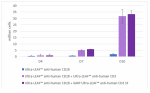
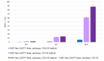
This data display is provided for general comparisons between formats.
Your actual data may vary due to variations in samples, target cells, instruments and their settings, staining conditions, and other factors.
If you need assistance with selecting the best format contact our expert technical support team.

Pricing & Availability
Clone
OKT3 (See other available formats)
Regulatory Status
RUO
Workshop
HCDM listed
Other Names
T3, CD3ε
Isotype
Mouse IgG2a, κ
Ave. Rating
Submit a Review
Product Citations
publications
OKT3_FITC_053107
Human peripheral blood lymphocytes stained with OKT3 FITC
  • OKT3_FITC_053107
    Human peripheral blood lymphocytes stained with OKT3 FITC
Compare all formats See FITC spectral data
Cat # Size Price Quantity Check Availability Save
317305 25 tests NOK231
Check Availability


Need larger quantities of this item?
Request Bulk Quote
317306 100 tests NOK454
Check Availability


Need larger quantities of this item?
Request Bulk Quote
Description

CD3ε is a 20 kD chain of the CD3/T cell receptor (TCR) complex, which is composed of two CD3ε, one CD3γ, one CD3δ, one CD3ζ (CD247), and a T cell receptor (α/β or γ/δ) heterodimer. It is found on all mature T lymphocytes, NK T cells, and some thymocytes. CD3, also known as T3, is a member of the immunoglobulin superfamily that plays a role in antigen recognition, signal transduction, and T cell activation.

Product Details
Technical Data Sheet (pdf)

Product Details

Verified Reactivity
Human
Antibody Type
Monoclonal
Host Species
Mouse
Formulation
Phosphate-buffered solution, pH 7.2, containing 0.09% sodium azide and BSA (origin USA)
Preparation
The antibody was purified by affinity chromatography, and conjugated with FITC under optimal conditions.
Concentration
Lot-specific (to obtain lot-specific concentration and expiration, please enter the lot number in our Certificate of Analysis online tool.)
Storage & Handling
The antibody solution should be stored undiluted between 2°C and 8°C, and protected from prolonged exposure to light. Do not freeze.
Application

FC - Quality tested

Recommended Usage

Each lot of this antibody is quality control tested by immunofluorescent staining with flow cytometric analysis. For flow cytometric staining, the suggested use of this reagent is 5 µl per million cells in 100 µl staining volume or 5 µl per 100 µl of whole blood.

Excitation Laser
Blue Laser (488 nm)
Application Notes

The OKT3 monoclonal antibody reacts with an epitope on the epsilon-subunit within the human CD3 complex.

Clone OKT3 can block the binding of clones SK7 and UCHT1.4 The OKT3 antibody is able to induce T cell activation. Additional reported applications (for the relevant formats) include: immunohistochemical staining of acetone-fixed frozen sections and activation of T cells. The LEAF™ purified antibody (Endotoxin <0.1 EU/µg, Azide-Free, 0.2 µm filtered) is recommended for functional assays (Cat. No. 317304). For highly sensitive assays, we recommend Ultra-LEAF™ purified antibody (Cat. No. 317326) with a lower endotoxin limit than standard LEAF™ purified antibodies (Endotoxin <0.01 EU/µg).

Application References
  1. Schlossman S, et al. Eds. 1995. Leucocyte Typing V. Oxford University Press. New York.
  2. Knapp W. 1989. Leucocyte Typing IV. Oxford University Press New York.
  3. Barclay N, et al. 1997. The Leucocyte Antigen Facts Book. Academic Press Inc. San Diego.
  4. Li B, et al. 2005. Immunology 116:487.
  5. Jeong HY, et al. 2008. J. Leuckocyte Biol. 83:755. PubMed
  6. Alter G, et al. 2008. J. Virol. 82:9668. PubMed
  7. Manevich-Mendelson E, et al. 2009. Blood 114:2344. PubMed
  8. Pinto JP, et al. 2010. Immunology. 130:217. PubMed
  9. Biggs MJ, et al. 2011. J. R. Soc. Interface. 8:1462. PubMed
Product Citations
  1. Gkika E, et al. 2023. NPJ Precis Oncol. 7:24. PubMed
  2. Li D, et al. 2023. Int J Oncol. 62:. PubMed
  3. Li L, et al. 2023. Heliyon. 9:e14219. PubMed
  4. Deng G, et al. 2023. Cancers (Basel). 15:. PubMed
  5. Kim JH, et al. 2020. Sci Rep. 10:1835. PubMed
  6. Zhang T, et al. 2022. Am J Reprod Immunol. 87:e13524. PubMed
  7. Tessaro FHG, et al. 2022. Cell Rep. 39:110977. PubMed
  8. Deepak HB, et al. 2022. Indian J Pharmacol. 54:183. PubMed
  9. Rosato F, et al. 2022. J Transl Med. 20:578. PubMed
  10. Zhang W, et al. 2022. Front Cell Dev Biol. 10:842813. PubMed
  11. Zitti B, et al. 2017. Sci Rep. 10.1038/s41598-017-10403-0. PubMed
  12. Afolabi LO, et al. 2021. Front Immunol. 12:701671. PubMed
  13. Guo X, et al. 2016. Mol Ther Methods Clin Dev. 3:15054. PubMed
  14. Want MY, et al. 2019. Oncoimmunology. 8:e1586042. PubMed
  15. Lugo-Reyes SO, et al. 2021. J Clin Immunol. 41:1291. PubMed
  16. Hess NJ, et al. 2020. J Immunol. 205:272. PubMed
  17. Clayton K, et al. 2014. J Immunol. 192:782. PubMed
  18. Catapano M, et al. 2020. J Invest Dermatol. 140:816. PubMed
  19. Shi Z, et al. 2021. Ann Clin Transl Neurol. 8:43. PubMed
  20. Wang Y, et al. 2019. J Transl Med. 17:93. PubMed
  21. Wu X, et al. 2020. Cancers (Basel). 12:. PubMed
  22. Moen IN, et al. 2021. Cell Death Discov. 7:36. PubMed
  23. Yamada E, et al. 2021. Gut Microbes. 13:1997292. PubMed
  24. Zhang B, et al. 2022. J Exp Med. 219:. PubMed
  25. Dai Q, et al. 2020. Front Immunol. 11:539654. PubMed
  26. Lin L, et al. 2018. Oncol Lett. 16:1259. PubMed
  27. Sun Y, et al. 2022. Nat Commun. 13:3916. PubMed
  28. Zhang Z, et al. 2021. Acta Pharm Sin B. 11:1965. PubMed
  29. Celik H, et al. 2021. Cancer Discov. 11:3126. PubMed
  30. Giesecke C, et al. 2017. Cytometry A. 10.1002/cyto.a.23250. PubMed
  31. Fujigaki J, et al. 2015. PLoS One. 10: 0132521. PubMed
  32. Gillissen MA, et al. 2018. Blood. 131:131. PubMed
  33. Kim K, et al. 2019. Nat Microbiol. 1.586111111. PubMed
  34. Sperk M, et al. 2021. iScience. 24:102111. PubMed
  35. Nakasone H, et al. 2021. Commun Biol. 4:1177. PubMed
  36. Wu X, et al. 2021. Front Immunol. 12:731767. PubMed
  37. Jeyanathan M, et al. 2022. JCI Insight. 7:. PubMed
  38. Omer OS, et al. 2020. Methods Mol Biol. 2121:199. PubMed
  39. James KR, et al. 2020. Nat Immunol. 1.113194444. PubMed
  40. Hazenberg MD, et al. 2019. Blood Adv. 2.659722222. PubMed
  41. Chulpanova DS, et al. 2021. Biology (Basel). 10:. PubMed
  42. Golebski K, et al. 2021. Immunity. 54(2):291-307.e7. PubMed
  43. Agashe VV, et al. 2018. J Immunol. 201:772. PubMed
  44. Oyer JL, et al. 2022. Front Immunol. 13:861681. PubMed
  45. Zhang CR, et al. 2022. Blood Cancer Discov. 3:220. PubMed
  46. Cheng J, et al. 2022. iScience. 25:103588. PubMed
  47. Tsai HI, et al. 2021. EMBO Mol Med. 13:e12834. PubMed
  48. Zhang W, et al. 2022. J Transl Med. 20:558. PubMed
  49. Aagaard K, et al. 2017. Sci Rep. 7:41389. PubMed
  50. Miyamae Y, et al. 2020. Cell Chemical Biology. 27(12):1573-1581.e3. PubMed
  51. Winkler C, et al. 2021. JCI Insight. 6:. PubMed
  52. Akusjärvi SS, et al. 2022. iScience. 25:103607. PubMed
  53. Zhou Z, et al. 2022. J Gastrointest Oncol. 13:732. PubMed
  54. Zhao B, et al. 2013. PLoS One. 8:77708. PubMed
  55. Bal S, et al. 2016. Nat Immunol. 10.1038/ni.3444. PubMed
  56. Gopalakrishnapillai A, et al. 2021. Cancers (Basel). 13:. PubMed
  57. Loo Yau H, et al. 2021. STAR Protocols. 2(2):100549. PubMed
  58. Shikhagaie M, et al. 2017. Cell Rep. 18(7):1761-1773. PubMed
  59. Sullivan KD et al. 2016. eLife. 5 pii: e16220. PubMed
  60. Ochi T, et al. 2011. Blood. 118:1495. PubMed
  61. Svensson Akusjärvi S, et al. 2022. Commun Biol. 5:357. PubMed
  62. Fierle JK, et al. 2021. Cell Reports Medicine. 2(8):100362. PubMed
  63. Gkika E, et al. 2023. NPJ Precis Oncol. 7:24. PubMed
  64. Li D, et al. 2023. Int J Oncol. 62:. PubMed
  65. Li L, et al. 2023. Heliyon. 9:e14219. PubMed
  66. Deng G, et al. 2023. Cancers (Basel). 15:. PubMed
  67. Kim JH, et al. 2020. Sci Rep. 10:1835. PubMed
  68. Zhang T, et al. 2022. Am J Reprod Immunol. 87:e13524. PubMed
  69. Tessaro FHG, et al. 2022. Cell Rep. 39:110977. PubMed
  70. Deepak HB, et al. 2022. Indian J Pharmacol. 54:183. PubMed
  71. Rosato F, et al. 2022. J Transl Med. 20:578. PubMed
  72. Zhang W, et al. 2022. Front Cell Dev Biol. 10:842813. PubMed
  73. Zitti B, et al. 2017. Sci Rep. 10.1038/s41598-017-10403-0. PubMed
  74. Afolabi LO, et al. 2021. Front Immunol. 12:701671. PubMed
  75. Guo X, et al. 2016. Mol Ther Methods Clin Dev. 3:15054. PubMed
  76. Want MY, et al. 2019. Oncoimmunology. 8:e1586042. PubMed
  77. Lugo-Reyes SO, et al. 2021. J Clin Immunol. 41:1291. PubMed
  78. Hess NJ, et al. 2020. J Immunol. 205:272. PubMed
  79. Clayton K, et al. 2014. J Immunol. 192:782. PubMed
  80. Catapano M, et al. 2020. J Invest Dermatol. 140:816. PubMed
  81. Shi Z, et al. 2021. Ann Clin Transl Neurol. 8:43. PubMed
  82. Wang Y, et al. 2019. J Transl Med. 17:93. PubMed
  83. Wu X, et al. 2020. Cancers (Basel). 12:. PubMed
  84. Moen IN, et al. 2021. Cell Death Discov. 7:36. PubMed
  85. Yamada E, et al. 2021. Gut Microbes. 13:1997292. PubMed
  86. Zhang B, et al. 2022. J Exp Med. 219:. PubMed
  87. Dai Q, et al. 2020. Front Immunol. 11:539654. PubMed
  88. Lin L, et al. 2018. Oncol Lett. 16:1259. PubMed
  89. Sun Y, et al. 2022. Nat Commun. 13:3916. PubMed
  90. Zhang Z, et al. 2021. Acta Pharm Sin B. 11:1965. PubMed
  91. Celik H, et al. 2021. Cancer Discov. 11:3126. PubMed
  92. Giesecke C, et al. 2017. Cytometry A. 10.1002/cyto.a.23250. PubMed
  93. Fujigaki J, et al. 2015. PLoS One. 10: 0132521. PubMed
  94. Gillissen MA, et al. 2018. Blood. 131:131. PubMed
  95. Kim K, et al. 2019. Nat Microbiol. 1.586111111. PubMed
  96. Sperk M, et al. 2021. iScience. 24:102111. PubMed
  97. Nakasone H, et al. 2021. Commun Biol. 4:1177. PubMed
  98. Wu X, et al. 2021. Front Immunol. 12:731767. PubMed
  99. Jeyanathan M, et al. 2022. JCI Insight. 7:. PubMed
  100. Omer OS, et al. 2020. Methods Mol Biol. 2121:199. PubMed
  101. James KR, et al. 2020. Nat Immunol. 1.113194444. PubMed
  102. Hazenberg MD, et al. 2019. Blood Adv. 2.659722222. PubMed
  103. Chulpanova DS, et al. 2021. Biology (Basel). 10:. PubMed
  104. Golebski K, et al. 2021. Immunity. 54(2):291-307.e7. PubMed
  105. Agashe VV, et al. 2018. J Immunol. 201:772. PubMed
  106. Oyer JL, et al. 2022. Front Immunol. 13:861681. PubMed
  107. Zhang CR, et al. 2022. Blood Cancer Discov. 3:220. PubMed
  108. Cheng J, et al. 2022. iScience. 25:103588. PubMed
  109. Tsai HI, et al. 2021. EMBO Mol Med. 13:e12834. PubMed
  110. Zhang W, et al. 2022. J Transl Med. 20:558. PubMed
  111. Aagaard K, et al. 2017. Sci Rep. 7:41389. PubMed
  112. Miyamae Y, et al. 2020. Cell Chemical Biology. 27(12):1573-1581.e3. PubMed
  113. Winkler C, et al. 2021. JCI Insight. 6:. PubMed
  114. Akusjärvi SS, et al. 2022. iScience. 25:103607. PubMed
  115. Zhou Z, et al. 2022. J Gastrointest Oncol. 13:732. PubMed
  116. Zhao B, et al. 2013. PLoS One. 8:77708. PubMed
  117. Bal S, et al. 2016. Nat Immunol. 10.1038/ni.3444. PubMed
  118. Gopalakrishnapillai A, et al. 2021. Cancers (Basel). 13:. PubMed
  119. Loo Yau H, et al. 2021. STAR Protocols. 2(2):100549. PubMed
  120. Shikhagaie M, et al. 2017. Cell Rep. 18(7):1761-1773. PubMed
  121. Sullivan KD et al. 2016. eLife. 5 pii: e16220. PubMed
  122. Ochi T, et al. 2011. Blood. 118:1495. PubMed
  123. Svensson Akusjärvi S, et al. 2022. Commun Biol. 5:357. PubMed
  124. Fierle JK, et al. 2021. Cell Reports Medicine. 2(8):100362. PubMed
RRID
AB_571906 (BioLegend Cat. No. 317305)
AB_571907 (BioLegend Cat. No. 317306)

Antigen Details

Structure
Ig superfamily, the subunits CD3γ, CD3δ, CD3ζ (CD247) and TCR (α/β or γ/δ) form the CD3/TCR complex, 20 kD
Distribution

Mature T and NK T cells, thymocyte differentiation

Function
Antigen recognition, signal transduction, T cell activation
Ligand/Receptor
Peptide antigen bound to MHC
Cell Type
NKT cells, T cells, Thymocytes, Tregs
Biology Area
Immunology
Molecular Family
CD Molecules
Antigen References
  1. Barclay N, et al. 1993. The Leucocyte FactsBook. Academic Press. San Diego.
  2. Beverly P, et al. 1981. Eur. J. Immunol. 11:329.
  3. Lanier L, et al. 1986. J. Immunol. 137:2501.
Gene ID
916 View all products for this Gene ID
UniProt
View information about CD3 on UniProt.org

Customers Also Purchased

Go To Top Version: 2    Revision Date: 06.12.2013

For Research Use Only. Not for diagnostic or therapeutic use.


This product is supplied subject to the terms and conditions, including the limited license, located at www.biolegend.com/terms) ("Terms") and may be used only as provided in the Terms. Without limiting the foregoing, BioLegend products may not be used for any Commercial Purpose as defined in the Terms, resold in any form, used in manufacturing, or reverse engineered, sequenced, or otherwise studied or used to learn its design or composition without express written approval of BioLegend. Regardless of the information given in this document, user is solely responsible for determining any license requirements necessary for user’s intended use and assumes all risk and liability arising from use of the product. BioLegend is not responsible for patent infringement or any other risks or liabilities whatsoever resulting from the use of its products.


BioLegend, the BioLegend logo, and all other trademarks are property of BioLegend, Inc. or their respective owners, and all rights are reserved.


8999 BioLegend Way, San Diego, CA 92121 www.biolegend.com
Toll-Free Phone: 1-877-Bio-Legend (246-5343) Phone: (858) 768-5800 Fax: (877) 455-9587

This data display is provided for general comparisons between formats.
Your actual data may vary due to variations in samples, target cells, instruments and their settings, staining conditions, and other factors.
If you need assistance with selecting the best format contact our expert technical support team.

ProductsHere

Login / Register
Remember me
Forgot your password? Reset password?
Create an Account