LEGEND MAX™ Mouse IFN-β ELISA Kit LEGEND MAX™ Mouse IFN-β ELISA Kit

Pricing & Availability
Regulatory Status
RUO
Other Names
Fibroblast interferon (Fi-IFN, F-IFN), Type-1 interferon, acid-stable interferon, R1-GI factor, IFN-β Pre-coated ELISA Kit
Ave. Rating
Submit a Review
Product Citations
publications
IFNb_Mouse_leg_070612
  • IFNb_Mouse_leg_070612
Cat # Size Price Quantity Check Availability Save
439407 1 Pre-coated Plate DKK3434
Check Availability


Need larger quantities of this item?
Request Bulk Quote
Description

Interferon-β is a member of the type I interferon class. Type I IFNs are involved in the innate immune system, being released non-specifically during the early stage of infection in response to stimuli such as viruses, dsRNA, LPS, and bacterial components. Once secreted by virus-infected or activated cells, they bind to the IFNα/β receptor (IFNAR), which is found on almost all cell types. Then, via the JAK-STAT pathway, they induce the expression of hundreds of IFN-stimulated genes (ISGs). While the function of many of these proteins remains to be elucidated, a hallmark of type I IFNs is their anti-viral activities, both general and specific against particular viruses. Type I IFNs also regulate cell growth and apoptosis, and exert various immunomodulatory and inflammatory effects. As such, they are clinically relevant, both as therapeutics and as targets of inhibition for the treatment of various autoimmune diseases and cancer.


Mouse IFN-β is a ~20 kD monomeric glycoprotein. It has 50% amino acid sequence homology with mouse IFN-α, its closest related protein, and 62% homology with human IFN-β. IFN-β is currently the therapeutic of choice for treating multiple sclerosis.

BioLegend’s LEGEND MAX™ Mouse IFN-β ELISA Kit is a sandwich Enzyme-Linked Immunosorbent Assay (ELISA), in which, a specific antibody is pre-coated on a 96-well strip-well plate.

BioLegend’s LEGEND MAX™ Mouse IFN-β ELISA Kit is specifically designed for the accurate quantitation of mouse IFN-β from cell culture supernatant, serum, plasma or other bodily fluids. It is ready-to-use, accurate, and sensitive.

Product Details
Technical Data Sheet (pdf)

Kit Details

Kit Contents
  • Anti-Mouse IFN-β Pre-coated 96-well Strip Microplate
  • Mouse IFN-β Dectection Antibody
  • Mouse IFN-β Standard
  • Avidin-HRP D
  • Matrix C (for serum and plasma samples only)
  • Assay Buffer A
  • Wash Buffer (20X)
  • Substrate Solution F
  • Stop Solution
  • Plate Sealers
Materials Not Included
  • Microplate reader able to measure absorbance at 450 nm
  • Adjustable pipettes to measure volumes ranging from 1 µL to 1,000 µL
  • Deionized water
  • Wash bottle or automated microplate washer
  • Log-Log graph paper or software for data analysis
  • Tubes to prepare standard dilutions
  • Timer
  • Plate Shaker
  • Polypropylene vials

Product Details

Verified Reactivity
Mouse
Application

ELISA

Application References
  1. Shah S, et al. 2013. J. Immunol. 191:3514. PubMed
  2. Wang S, et al. 2013. J Virol. 87:11851. PubMed
Product Citations
  1. Casazza RL, et al. 2022. MBio. 13:e0385721. PubMed
  2. Wang Y, et al. 2022. Cell Rep. 41:111434. PubMed
  3. Park JH, et al. 2022. Cell Rep. 41:111594. PubMed
  4. Wei K, et al. 2023. Signal Transduct Target Ther. 8:22. PubMed
  5. Xie X, et al. 2021. BMC Med. 19:247. PubMed
  6. Oldenburg R, et al. 2018. Front Immunol. 9:2. PubMed
  7. Zhou C, et al. 2020. Immunity. 52:767. PubMed
  8. Shah S, et al. 2013. J Immunol. 191:3514. PubMed
  9. Yan S, et al. 2018. J Immunol. 200:1016. PubMed
  10. Zhao M, et al. 2021. PLoS Pathog. 17:e1009901. PubMed
  11. Klopfenstein N, et al. 2021. PLoS Pathog. 17:e1009387. PubMed
  12. Zhou P, et al. 2018. Nat Commun. 9:1243. PubMed
  13. Cheng WY, et al. 2018. Front Immunol. 9:1297. PubMed
  14. Yoshida A, et al. 2018. J Virol. 92:e02094. PubMed
  15. Wu D, et al. 2022. Cell Death Dis. 13:731. PubMed
  16. Piersma SJ, et al. 2020. Elife. 9:00. PubMed
  17. Jiang H, et al. 2016. J Immunol. 197: 2880 - 2890. PubMed
  18. Kwon HK, et al. 2019. Exp Mol Med. 51:50. PubMed
  19. Fritzlar S, et al. 2019. MBio. 10:e00960. PubMed
  20. Ferri F, et al. 2015. Nat Commun. 6: 8900. PubMed
  21. Synn CB, et al. 2022. Yonsei Med J. 63:42. PubMed
  22. Chen Y, et al. 2021. Front Immunol. 12:672165. PubMed
  23. Li K, et al. 2021. Sci Adv. 7: . PubMed
  24. Stavrou S, et al. 2018. MBio. 9:e00923. PubMed
  25. Srikanth S, et al. 2019. Nat Immunol. 20:152. PubMed
  26. Kim KS, et al. 2021. J Leukoc Biol. 109:865. PubMed
  27. Reimer E, et al. 2020. J Cell Sci. 134:00:00. PubMed
  28. Nanda S, et al. 2016. J Immunol. 197(11):4266-4273. PubMed
  29. Sousa J, et al. 2020. Nat Commun. 11:1949. PubMed
  30. Shepardson K, et al. 2016. MBio. 7: e00506-16. PubMed
  31. Wu Y, Ren D, Chen G 2016. J Immunol. 197: 3336 - 3347. PubMed
  32. Zhu M, et al. 2015. J Immunol. 195: 289 - 297. PubMed
  33. Huang F, et al. 2021. Front Immunol. 12:707298. PubMed
  34. Raychowdhury R, et al. 2021. Immunohorizons. 5:818. PubMed
  35. Chen X, et al. 2021. J Immunol. :. PubMed
  36. Qiu W, et al. 2021. Nat Commun. 1582:12. PubMed
  37. Khan M, et al. 2020. PLoS Pathog. 16:e1009020. PubMed
  38. Lv M, et al. 2015. Sci Rep. 5: 12954. PubMed
  39. Wang L, et al. 2022. Nat Commun. 13:7107. PubMed
  40. Li X, et al. 2022. Sci Rep. 12:17058. PubMed
  41. Zhang Y, et al. 2022. Nat Commun. 13:4948. PubMed
  42. Saddawi-Konefka R, et al. 2022. Nat Commun. 13:4298. PubMed
  43. Yang S, et al. 2019. Sci Adv. 5:eaav7999. PubMed
  44. Karki R, et al. 2020. J Immunol. 204:2514. PubMed
  45. Wang S, et al. 2013. J Virol. 87:11851. PubMed
  46. Tong Y, et al. 2018. EBioMedicine. 39:132. PubMed
  47. Casazza RL, et al. 2022. MBio. 13:e0385721. PubMed
  48. Wang Y, et al. 2022. Cell Rep. 41:111434. PubMed
  49. Park JH, et al. 2022. Cell Rep. 41:111594. PubMed
  50. Wei K, et al. 2023. Signal Transduct Target Ther. 8:22. PubMed
  51. Xie X, et al. 2021. BMC Med. 19:247. PubMed
  52. Oldenburg R, et al. 2018. Front Immunol. 9:2. PubMed
  53. Zhou C, et al. 2020. Immunity. 52:767. PubMed
  54. Shah S, et al. 2013. J Immunol. 191:3514. PubMed
  55. Yan S, et al. 2018. J Immunol. 200:1016. PubMed
  56. Zhao M, et al. 2021. PLoS Pathog. 17:e1009901. PubMed
  57. Klopfenstein N, et al. 2021. PLoS Pathog. 17:e1009387. PubMed
  58. Zhou P, et al. 2018. Nat Commun. 9:1243. PubMed
  59. Cheng WY, et al. 2018. Front Immunol. 9:1297. PubMed
  60. Yoshida A, et al. 2018. J Virol. 92:e02094. PubMed
  61. Wu D, et al. 2022. Cell Death Dis. 13:731. PubMed
  62. Piersma SJ, et al. 2020. Elife. 9:00. PubMed
  63. Jiang H, et al. 2016. J Immunol. 197: 2880 - 2890. PubMed
  64. Kwon HK, et al. 2019. Exp Mol Med. 51:50. PubMed
  65. Fritzlar S, et al. 2019. MBio. 10:e00960. PubMed
  66. Ferri F, et al. 2015. Nat Commun. 6: 8900. PubMed
  67. Synn CB, et al. 2022. Yonsei Med J. 63:42. PubMed
  68. Chen Y, et al. 2021. Front Immunol. 12:672165. PubMed
  69. Li K, et al. 2021. Sci Adv. 7: . PubMed
  70. Stavrou S, et al. 2018. MBio. 9:e00923. PubMed
  71. Srikanth S, et al. 2019. Nat Immunol. 20:152. PubMed
  72. Kim KS, et al. 2021. J Leukoc Biol. 109:865. PubMed
  73. Reimer E, et al. 2020. J Cell Sci. 134:00:00. PubMed
  74. Nanda S, et al. 2016. J Immunol. 197(11):4266-4273. PubMed
  75. Sousa J, et al. 2020. Nat Commun. 11:1949. PubMed
  76. Shepardson K, et al. 2016. MBio. 7: e00506-16. PubMed
  77. Wu Y, Ren D, Chen G 2016. J Immunol. 197: 3336 - 3347. PubMed
  78. Zhu M, et al. 2015. J Immunol. 195: 289 - 297. PubMed
  79. Huang F, et al. 2021. Front Immunol. 12:707298. PubMed
  80. Raychowdhury R, et al. 2021. Immunohorizons. 5:818. PubMed
  81. Chen X, et al. 2021. J Immunol. :. PubMed
  82. Qiu W, et al. 2021. Nat Commun. 1582:12. PubMed
  83. Khan M, et al. 2020. PLoS Pathog. 16:e1009020. PubMed
  84. Lv M, et al. 2015. Sci Rep. 5: 12954. PubMed
  85. Wang L, et al. 2022. Nat Commun. 13:7107. PubMed
  86. Li X, et al. 2022. Sci Rep. 12:17058. PubMed
  87. Zhang Y, et al. 2022. Nat Commun. 13:4948. PubMed
  88. Saddawi-Konefka R, et al. 2022. Nat Commun. 13:4298. PubMed
  89. Yang S, et al. 2019. Sci Adv. 5:eaav7999. PubMed
  90. Karki R, et al. 2020. J Immunol. 204:2514. PubMed
  91. Wang S, et al. 2013. J Virol. 87:11851. PubMed
  92. Tong Y, et al. 2018. EBioMedicine. 39:132. PubMed
Sensitivity
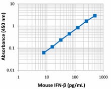
1.9 pg/mL
Standard Range
7.8-500 pg/mL

Antigen Details

Cell Sources
Fibroblasts, epithelial cells
Biology Area
Apoptosis/Tumor Suppressors/Cell Death, Cell Biology, Signal Transduction
Molecular Family
Cytokines/Chemokines
Gene ID
15977 View all products for this Gene ID
UniProt
View information about IFN-beta on UniProt.org

Related FAQs

In your LEGEND MAX™ ELISA Kits, there is a step that calls for washing the plates before adding sample. What is the purpose of this step?

We typically use a stabilizer for pre-coated plates. The additional washing step is designed to remove these components before you start the assay. If you do not perform the washing, the effect on assay performance is negligible.

I have multiple LEGEND MAX™ ELISA kits that I want to run simultaneously. Can I use the same wash buffer for all the kits?

The wash buffer provided in all our LEGEND MAX™ kits is the same and the part numbers on the wash buffer bottles in these kits should be identical. For ELISA MAX™ Deluxe and ELISA MAX™ Standard Sets, we provide a recipe for the wash buffer on each kit’s technical data sheet. This recipe is the same for all ELISA MAX™ sets.

For some of your ELISA kits, why do my serum samples require dilution with assay buffer?

In some cases, dilution with assay buffer is required to minimize the matrix difference between the samples and the standards to achieve better accuracy.

Go To Top Version: 4    Revision Date: 01/16/2017

For Research Use Only. Not for diagnostic or therapeutic use.


This product is supplied subject to the terms and conditions, including the limited license, located at www.biolegend.com/terms) ("Terms") and may be used only as provided in the Terms. Without limiting the foregoing, BioLegend products may not be used for any Commercial Purpose as defined in the Terms, resold in any form, used in manufacturing, or reverse engineered, sequenced, or otherwise studied or used to learn its design or composition without express written approval of BioLegend. Regardless of the information given in this document, user is solely responsible for determining any license requirements necessary for user’s intended use and assumes all risk and liability arising from use of the product. BioLegend is not responsible for patent infringement or any other risks or liabilities whatsoever resulting from the use of its products.


BioLegend, the BioLegend logo, and all other trademarks are property of BioLegend, Inc. or their respective owners, and all rights are reserved.


8999 BioLegend Way, San Diego, CA 92121 www.biolegend.com
Toll-Free Phone: 1-877-Bio-Legend (246-5343) Phone: (858) 768-5800 Fax: (877) 455-9587

Pricing & Availability
Regulatory Status
RUO
Other Names
Fibroblast interferon (Fi-IFN, F-IFN), Type-1 interferon, acid-stable interferon, R1-GI factor, IFN-β Pre-coated ELISA Kit
Ave. Rating
Submit a Review
Product Citations
publications
IFNb_Mouse_leg_070612
  • IFNb_Mouse_leg_070612
Cat # Size Price Quantity Check Availability Save
439407 1 Pre-coated Plate DKK3434
Check Availability


Need larger quantities of this item?
Request Bulk Quote
Description

Interferon-β is a member of the type I interferon class. Type I IFNs are involved in the innate immune system, being released non-specifically during the early stage of infection in response to stimuli such as viruses, dsRNA, LPS, and bacterial components. Once secreted by virus-infected or activated cells, they bind to the IFNα/β receptor (IFNAR), which is found on almost all cell types. Then, via the JAK-STAT pathway, they induce the expression of hundreds of IFN-stimulated genes (ISGs). While the function of many of these proteins remains to be elucidated, a hallmark of type I IFNs is their anti-viral activities, both general and specific against particular viruses. Type I IFNs also regulate cell growth and apoptosis, and exert various immunomodulatory and inflammatory effects. As such, they are clinically relevant, both as therapeutics and as targets of inhibition for the treatment of various autoimmune diseases and cancer.


Mouse IFN-β is a ~20 kD monomeric glycoprotein. It has 50% amino acid sequence homology with mouse IFN-α, its closest related protein, and 62% homology with human IFN-β. IFN-β is currently the therapeutic of choice for treating multiple sclerosis.

BioLegend’s LEGEND MAX™ Mouse IFN-β ELISA Kit is a sandwich Enzyme-Linked Immunosorbent Assay (ELISA), in which, a specific antibody is pre-coated on a 96-well strip-well plate.

BioLegend’s LEGEND MAX™ Mouse IFN-β ELISA Kit is specifically designed for the accurate quantitation of mouse IFN-β from cell culture supernatant, serum, plasma or other bodily fluids. It is ready-to-use, accurate, and sensitive.

Product Details
Technical Data Sheet (pdf)

Kit Details

Kit Contents
  • Anti-Mouse IFN-β Pre-coated 96-well Strip Microplate
  • Mouse IFN-β Dectection Antibody
  • Mouse IFN-β Standard
  • Avidin-HRP D
  • Matrix C (for serum and plasma samples only)
  • Assay Buffer A
  • Wash Buffer (20X)
  • Substrate Solution F
  • Stop Solution
  • Plate Sealers
Materials Not Included
  • Microplate reader able to measure absorbance at 450 nm
  • Adjustable pipettes to measure volumes ranging from 1 µL to 1,000 µL
  • Deionized water
  • Wash bottle or automated microplate washer
  • Log-Log graph paper or software for data analysis
  • Tubes to prepare standard dilutions
  • Timer
  • Plate Shaker
  • Polypropylene vials

Product Details

Verified Reactivity
Mouse
Application

ELISA

Application References
  1. Shah S, et al. 2013. J. Immunol. 191:3514. PubMed
  2. Wang S, et al. 2013. J Virol. 87:11851. PubMed
Product Citations
  1. Casazza RL, et al. 2022. MBio. 13:e0385721. PubMed
  2. Wang Y, et al. 2022. Cell Rep. 41:111434. PubMed
  3. Park JH, et al. 2022. Cell Rep. 41:111594. PubMed
  4. Wei K, et al. 2023. Signal Transduct Target Ther. 8:22. PubMed
  5. Xie X, et al. 2021. BMC Med. 19:247. PubMed
  6. Oldenburg R, et al. 2018. Front Immunol. 9:2. PubMed
  7. Zhou C, et al. 2020. Immunity. 52:767. PubMed
  8. Shah S, et al. 2013. J Immunol. 191:3514. PubMed
  9. Yan S, et al. 2018. J Immunol. 200:1016. PubMed
  10. Zhao M, et al. 2021. PLoS Pathog. 17:e1009901. PubMed
  11. Klopfenstein N, et al. 2021. PLoS Pathog. 17:e1009387. PubMed
  12. Zhou P, et al. 2018. Nat Commun. 9:1243. PubMed
  13. Cheng WY, et al. 2018. Front Immunol. 9:1297. PubMed
  14. Yoshida A, et al. 2018. J Virol. 92:e02094. PubMed
  15. Wu D, et al. 2022. Cell Death Dis. 13:731. PubMed
  16. Piersma SJ, et al. 2020. Elife. 9:00. PubMed
  17. Jiang H, et al. 2016. J Immunol. 197: 2880 - 2890. PubMed
  18. Kwon HK, et al. 2019. Exp Mol Med. 51:50. PubMed
  19. Fritzlar S, et al. 2019. MBio. 10:e00960. PubMed
  20. Ferri F, et al. 2015. Nat Commun. 6: 8900. PubMed
  21. Synn CB, et al. 2022. Yonsei Med J. 63:42. PubMed
  22. Chen Y, et al. 2021. Front Immunol. 12:672165. PubMed
  23. Li K, et al. 2021. Sci Adv. 7: . PubMed
  24. Stavrou S, et al. 2018. MBio. 9:e00923. PubMed
  25. Srikanth S, et al. 2019. Nat Immunol. 20:152. PubMed
  26. Kim KS, et al. 2021. J Leukoc Biol. 109:865. PubMed
  27. Reimer E, et al. 2020. J Cell Sci. 134:00:00. PubMed
  28. Nanda S, et al. 2016. J Immunol. 197(11):4266-4273. PubMed
  29. Sousa J, et al. 2020. Nat Commun. 11:1949. PubMed
  30. Shepardson K, et al. 2016. MBio. 7: e00506-16. PubMed
  31. Wu Y, Ren D, Chen G 2016. J Immunol. 197: 3336 - 3347. PubMed
  32. Zhu M, et al. 2015. J Immunol. 195: 289 - 297. PubMed
  33. Huang F, et al. 2021. Front Immunol. 12:707298. PubMed
  34. Raychowdhury R, et al. 2021. Immunohorizons. 5:818. PubMed
  35. Chen X, et al. 2021. J Immunol. :. PubMed
  36. Qiu W, et al. 2021. Nat Commun. 1582:12. PubMed
  37. Khan M, et al. 2020. PLoS Pathog. 16:e1009020. PubMed
  38. Lv M, et al. 2015. Sci Rep. 5: 12954. PubMed
  39. Wang L, et al. 2022. Nat Commun. 13:7107. PubMed
  40. Li X, et al. 2022. Sci Rep. 12:17058. PubMed
  41. Zhang Y, et al. 2022. Nat Commun. 13:4948. PubMed
  42. Saddawi-Konefka R, et al. 2022. Nat Commun. 13:4298. PubMed
  43. Yang S, et al. 2019. Sci Adv. 5:eaav7999. PubMed
  44. Karki R, et al. 2020. J Immunol. 204:2514. PubMed
  45. Wang S, et al. 2013. J Virol. 87:11851. PubMed
  46. Tong Y, et al. 2018. EBioMedicine. 39:132. PubMed
  47. Casazza RL, et al. 2022. MBio. 13:e0385721. PubMed
  48. Wang Y, et al. 2022. Cell Rep. 41:111434. PubMed
  49. Park JH, et al. 2022. Cell Rep. 41:111594. PubMed
  50. Wei K, et al. 2023. Signal Transduct Target Ther. 8:22. PubMed
  51. Xie X, et al. 2021. BMC Med. 19:247. PubMed
  52. Oldenburg R, et al. 2018. Front Immunol. 9:2. PubMed
  53. Zhou C, et al. 2020. Immunity. 52:767. PubMed
  54. Shah S, et al. 2013. J Immunol. 191:3514. PubMed
  55. Yan S, et al. 2018. J Immunol. 200:1016. PubMed
  56. Zhao M, et al. 2021. PLoS Pathog. 17:e1009901. PubMed
  57. Klopfenstein N, et al. 2021. PLoS Pathog. 17:e1009387. PubMed
  58. Zhou P, et al. 2018. Nat Commun. 9:1243. PubMed
  59. Cheng WY, et al. 2018. Front Immunol. 9:1297. PubMed
  60. Yoshida A, et al. 2018. J Virol. 92:e02094. PubMed
  61. Wu D, et al. 2022. Cell Death Dis. 13:731. PubMed
  62. Piersma SJ, et al. 2020. Elife. 9:00. PubMed
  63. Jiang H, et al. 2016. J Immunol. 197: 2880 - 2890. PubMed
  64. Kwon HK, et al. 2019. Exp Mol Med. 51:50. PubMed
  65. Fritzlar S, et al. 2019. MBio. 10:e00960. PubMed
  66. Ferri F, et al. 2015. Nat Commun. 6: 8900. PubMed
  67. Synn CB, et al. 2022. Yonsei Med J. 63:42. PubMed
  68. Chen Y, et al. 2021. Front Immunol. 12:672165. PubMed
  69. Li K, et al. 2021. Sci Adv. 7: . PubMed
  70. Stavrou S, et al. 2018. MBio. 9:e00923. PubMed
  71. Srikanth S, et al. 2019. Nat Immunol. 20:152. PubMed
  72. Kim KS, et al. 2021. J Leukoc Biol. 109:865. PubMed
  73. Reimer E, et al. 2020. J Cell Sci. 134:00:00. PubMed
  74. Nanda S, et al. 2016. J Immunol. 197(11):4266-4273. PubMed
  75. Sousa J, et al. 2020. Nat Commun. 11:1949. PubMed
  76. Shepardson K, et al. 2016. MBio. 7: e00506-16. PubMed
  77. Wu Y, Ren D, Chen G 2016. J Immunol. 197: 3336 - 3347. PubMed
  78. Zhu M, et al. 2015. J Immunol. 195: 289 - 297. PubMed
  79. Huang F, et al. 2021. Front Immunol. 12:707298. PubMed
  80. Raychowdhury R, et al. 2021. Immunohorizons. 5:818. PubMed
  81. Chen X, et al. 2021. J Immunol. :. PubMed
  82. Qiu W, et al. 2021. Nat Commun. 1582:12. PubMed
  83. Khan M, et al. 2020. PLoS Pathog. 16:e1009020. PubMed
  84. Lv M, et al. 2015. Sci Rep. 5: 12954. PubMed
  85. Wang L, et al. 2022. Nat Commun. 13:7107. PubMed
  86. Li X, et al. 2022. Sci Rep. 12:17058. PubMed
  87. Zhang Y, et al. 2022. Nat Commun. 13:4948. PubMed
  88. Saddawi-Konefka R, et al. 2022. Nat Commun. 13:4298. PubMed
  89. Yang S, et al. 2019. Sci Adv. 5:eaav7999. PubMed
  90. Karki R, et al. 2020. J Immunol. 204:2514. PubMed
  91. Wang S, et al. 2013. J Virol. 87:11851. PubMed
  92. Tong Y, et al. 2018. EBioMedicine. 39:132. PubMed
Sensitivity
1.9 pg/mL
Standard Range
7.8-500 pg/mL

Antigen Details

Cell Sources
Fibroblasts, epithelial cells
Biology Area
Apoptosis/Tumor Suppressors/Cell Death, Cell Biology, Signal Transduction
Molecular Family
Cytokines/Chemokines
Gene ID
15977 View all products for this Gene ID
UniProt
View information about IFN-beta on UniProt.org

Related FAQs

In your LEGEND MAX™ ELISA Kits, there is a step that calls for washing the plates before adding sample. What is the purpose of this step?

We typically use a stabilizer for pre-coated plates. The additional washing step is designed to remove these components before you start the assay. If you do not perform the washing, the effect on assay performance is negligible.

I have multiple LEGEND MAX™ ELISA kits that I want to run simultaneously. Can I use the same wash buffer for all the kits?

The wash buffer provided in all our LEGEND MAX™ kits is the same and the part numbers on the wash buffer bottles in these kits should be identical. For ELISA MAX™ Deluxe and ELISA MAX™ Standard Sets, we provide a recipe for the wash buffer on each kit’s technical data sheet. This recipe is the same for all ELISA MAX™ sets.

For some of your ELISA kits, why do my serum samples require dilution with assay buffer?

In some cases, dilution with assay buffer is required to minimize the matrix difference between the samples and the standards to achieve better accuracy.

Go To Top Version: 4    Revision Date: 01/16/2017

For Research Use Only. Not for diagnostic or therapeutic use.


This product is supplied subject to the terms and conditions, including the limited license, located at www.biolegend.com/terms) ("Terms") and may be used only as provided in the Terms. Without limiting the foregoing, BioLegend products may not be used for any Commercial Purpose as defined in the Terms, resold in any form, used in manufacturing, or reverse engineered, sequenced, or otherwise studied or used to learn its design or composition without express written approval of BioLegend. Regardless of the information given in this document, user is solely responsible for determining any license requirements necessary for user’s intended use and assumes all risk and liability arising from use of the product. BioLegend is not responsible for patent infringement or any other risks or liabilities whatsoever resulting from the use of its products.


BioLegend, the BioLegend logo, and all other trademarks are property of BioLegend, Inc. or their respective owners, and all rights are reserved.


8999 BioLegend Way, San Diego, CA 92121 www.biolegend.com
Toll-Free Phone: 1-877-Bio-Legend (246-5343) Phone: (858) 768-5800 Fax: (877) 455-9587

ProductsHere

Login / Register
Remember me
Forgot your password? Reset password?
Create an Account