Alexa Fluor® 488 anti-mouse CD45 Antibody Alexa Fluor® 488 anti-mouse CD45 Antibody

Pricing & Availability
Clone
30-F11 (See other available formats)
Regulatory Status
RUO
Other Names
T200, Ly-5, LCA
Isotype
Rat IgG2b, κ
Ave. Rating
Submit a Review
Product Citations
publications
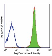
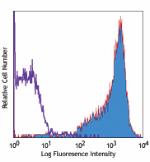
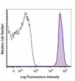
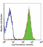
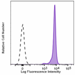
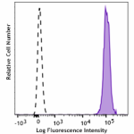
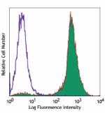
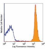
30-F11_Alx488_090507
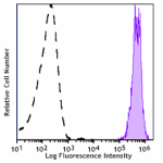
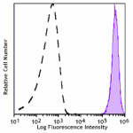
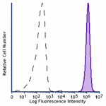
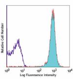
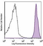
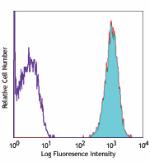
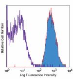
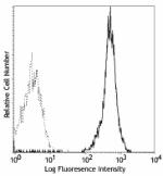
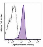
C57BL/6 mouse splenocytes stained with 30-F11 Alexa Fluor® 488
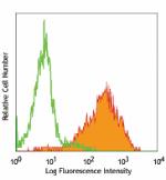
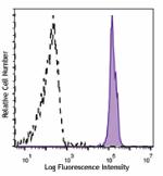
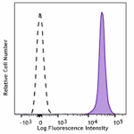
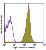
  • 30-F11_Alx488_090507
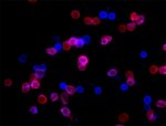
    C57BL/6 mouse splenocytes stained with 30-F11 Alexa Fluor® 488
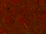
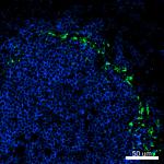
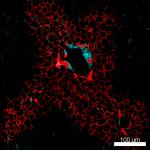
  • 33_Mouse_Spleen_CD169_CD45
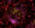
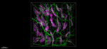
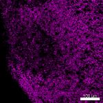
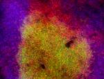
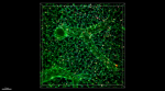
    Confocal image of C57BL/6 mouse spleen sample acquired using the IBEX method of highly multiplexed antibody-based imaging: CD169 (green) in Cycle 2 and CD45 (blue) in Cycle 3. Tissues were prepared using ~1% (vol/vol) formaldehyde and a detergent. Following fixation, samples are immersed in 30% (wt/vol) sucrose for cryoprotection. Images are courtesy of Drs. Andrea J. Radtke and Ronald N. Germain of the Center for Advanced Tissue Imaging (CAT-I) in the National Institute of Allergy and Infectious Diseases (NIAID, NIH).
Compare all formats See Alexa Fluor® 488 spectral data
Cat # Size Price Quantity Check Availability Save
103122 100 µg NOK1890
Check Availability


Need larger quantities of this item?
Request Bulk Quote
103121 25 µg NOK834
Check Availability


Need larger quantities of this item?
Request Bulk Quote
Description

CD45 is a 180-240 kD glycoprotein also known as the leukocyte common antigen (LCA), T200, or Ly-5. It is a member of the protein tyrosine phosphatase (PTP) family, expressed on all hematopoietic cells except mature erythrocytes and platelets. There are different isoforms of CD45 that arise from variable splicing of exons 4, 5, and 6, which encode A, B, and C determinants, respectively. CD45 plays a key role in TCR and BCR signal transduction. These isoforms are very specific to the activation and maturation state of the cell as well as cell type. The primary ligands for CD45 are galectin-1, CD2, CD3, CD4, TCR, CD22, and Thy-1.

Product Details
Technical Data Sheet (pdf)

Product Details

Verified Reactivity
Mouse
Antibody Type
Monoclonal
Host Species
Rat
Immunogen
Mouse thymus or spleen
Formulation
Phosphate-buffered solution, pH 7.2, containing 0.09% sodium azide.
Preparation
The antibody was purified by affinity chromatography and conjugated with Alexa Fluor® 488 under optimal conditions.
Concentration
0.5 mg/mL
Storage & Handling
The antibody solution should be stored undiluted between 2°C and 8°C, and protected from prolonged exposure to light. Do not freeze.
Application

FC - Quality tested

SB - Reported in the literature, not verified in house

Recommended Usage

Each lot of this antibody is quality control tested by immunofluorescent staining with flow cytometric analysis. For flow cytometric staining, the suggested use of this reagent is ≤ 0.25 µg per 106 cells in 100 µL volume. It is recommended that the reagent be titrated for optimal performance for each application.

* Alexa Fluor® 488 has a maximum emission of 519 nm when it is excited at 488 nm.


Alexa Fluor® and Pacific Blue™ are trademarks of Life Technologies Corporation.

View full statement regarding label licenses
Excitation Laser
Blue Laser (488 nm)
Application Notes

Clone 30-F11 reacts with all isoforms and both CD45.1 and CD45.2 alloantigens of CD45.

Additional reported applications (for relevant formats) include: immunoprecipitation3, complement-dependent cytotoxicity1,5, immunohistochemistry (acetone-fixed frozen sections, zinc-fixed paraffin-embedded sections and formalin-fixed paraffin-embedded sections)4,6, Western blotting7, and spatial biology (IBEX)10,11. The Ultra-LEAF™ purified antibody (Endotoxin < 0.01 EU/µg, Azide-Free, 0.2 µm filtered) is recommended for functional assays (Cat. No. 103163 and 103164).

Additional Product Notes

Iterative Bleaching Extended multi-pleXity (IBEX) is a fluorescent imaging technique capable of highly-multiplexed spatial analysis. The method relies on cyclical bleaching of panels of fluorescent antibodies in order to image and analyze many markers over multiple cycles of staining, imaging, and, bleaching. It is a community-developed open-access method developed by the Center for Advanced Tissue Imaging (CAT-I) in the National Institute of Allergy and Infectious Diseases (NIAID, NIH).

Application References
  1. Podd BS, et al. 2006. J. Immunol. 176:6532. (FC, CMCD) PubMed
  2. Haynes NM, et al. 2007. J. Immunol. 179:5099. (FC)
  3. Ledbetter JA, et al. 1979. Immunol. Rev. 47:63. (IP)
  4. Simon DI, et al. 2000. J. Clin. Invest. 105:293. (IHC)
  5. Seaman WE. 1983. J. Immunol. 130:1713. (CMCD)
  6. Cornet A, et al. 2001. P. Natl. Acad. Sci. USA 98:13306. (IHC)
  7. Tsuboi S and Fukuda M. 1998. J. Biol. Chem. 273:30680. (WB) PubMed
  8. Liu F, et al. 2012. Blood. 119:3295. PubMed
  9. Pelletier AN, et al. 2012. J. Immunol. 188:5561. PubMed
  10. Radtke AJ, et al. 2020. Proc Natl Acad Sci U S A. 117:33455-65. (SB) PubMed
  11. Radtke AJ, et al. 2022. Nat Protoc. 17:378-401. (SB) PubMed
Product Citations
  1. Preda MB, et al. 2021. Cell Death Dis. 12:566. PubMed
  2. Kuo PC, et al. 2021. Brain Commun. 3:fcab187. PubMed
  3. Wu Y, et al. 2021. Immunity. 54:2595. PubMed
  4. Wemlinger SM, et al. 2022. J Immunol. 208:1566. PubMed
  5. Liu H, et al. 2022. Cell Rep Med. 3:100660. PubMed
  6. Frederico B, et al. 2022. Dev Cell. 57:1957. PubMed
  7. Ullah I, et al. 2023. Cell Rep Med. 4:100893. PubMed
  8. Olivares-González L, et al. 2022. Antioxidants (Basel). 12: . PubMed
  9. Gilman KE, et al. 2023. Front Immunol. 13:1045710. PubMed
  10. Parida PK, et al. 2022. Cell Metab. 34:90. PubMed
  11. Gomez-Salinero JM, et al. 2022. Cell Stem Cell. 29:593. PubMed
  12. Luff SA, et al. 2022. Nat Cell Biol. 24:616. PubMed
  13. Battis K, et al. 2022. J Neurosci. 42:7673. PubMed
  14. Abdel-Haq R, et al. 2022. Elife. 11:. PubMed
  15. Axelrod ML, et al. 2022. Nature. 611:818. PubMed
  16. Gómez-Salinero JM, et al. 2022. Nat Cardiovasc Res. 1:882. PubMed
  17. Fan Z, et al. 2023. Angiogenesis. . PubMed
  18. Gilman KE, et al. 2023. Cancers (Basel). 15:. PubMed
  19. Palakurthi B, et al. 2023. Nat Commun. 14:2109. PubMed
  20. Fixsen BR, et al. 2023. Nat Immunol. . PubMed
  21. Marko C, et al. 2014. Invest Ophthalmol Vis Sci . 55:291. PubMed
  22. Giorgetti E, et al. 2020. Cell Reports. 29(6):1539-1554.e7.. PubMed
  23. Kazakevych J, et al. 2020. Genome Biol. 21:64. PubMed
  24. Sakai M, et al. 2020. Immunity. 51(4):655-670. PubMed
  25. Schmidleithner L et al. 2019. Immunity. 50(5):1232-1248 . PubMed
  26. Logan C, Bowen C, and Menko A 2017. Sci Rep. . 10.1038/s41598-017-16456-5. PubMed
  27. Petursdottir D, et al. 2017. Front Immunol. . 10.3389/fimmu.2017.01699. PubMed
  28. Nagai Y, et al. 2019. Front Immunol. 10:174. PubMed
  29. Khandelwal P, et al. 2013. PLoS One. 8:64193. PubMed
  30. Masschelein E, et al. 2020. Skelet Muscle. 10:21. PubMed
  31. Martínez-Sabadell A, et al. 2022. STAR Protoc. 3:101712. PubMed
  32. Rodriguez AB, et al. 2021. Cell Reports. 36(3):109422. PubMed
  33. Bornes L, et al. 2021. Life Sci Alliance. 4:00. PubMed
  34. Jenkins RW, et al. 2018. Cancer Discov. 8:196. PubMed
  35. Dubey LK, et al. 2019. Cell Rep. 27:2442. PubMed
  36. DeDreu J, et al. 2020. FASEB J. 34:9316. PubMed
  37. Dos Santos Dias L, et al. 2021. PLoS Pathog. e1009324:17. PubMed
  38. Hallett JM, et al. 2022. Cell Stem Cell. 29:355. PubMed
  39. Mehta AK, et al. 2021. Nat Cancer. 2:66. PubMed
  40. Yip HYK, et al. 2021. STAR Protocols. 2(3):100765. PubMed
  41. Chen I, et al. 2016. Sci Rep. 6:27195. PubMed
  42. Duarte J, et al. 2016. J Immunol. 197(12):4838-4847. PubMed
  43. Jung Y, et al. 2021. Nat Commun. 12:3872. PubMed
  44. Scarneo SA, et al. 2022. Sci Rep. 12:18091. PubMed
  45. Gutknecht M, et al. 2017. Nat Commun. 10.1038/s41467-017-00488-6. PubMed
  46. Ferrari G, et al. 2011. Invest Ophthalmol Vis Sci. 52:2532. PubMed
  47. Mizbani A, et al. 2016. Development. 143: 4137 - 4148. PubMed
  48. Burns JC, et al. 2020. eLife. 9:00. PubMed
  49. Wara AK, et al. 2020. Cell Rep. 33:108550. PubMed
  50. Wu H, et al. 2021. Cell Death Discov. 7:225. PubMed
  51. Lustgarten Guahmich N, et al. 2020. Angiogenesis. 23:443. PubMed
  52. Limbad C, et al. 2022. iScience. 25:103848. PubMed
  53. Yip HYK, et al. 2020. Molecular Cell. 80(2):279-295.e8. PubMed
  54. Liu X, et al. 2020. Cell Host Microbe. 28(5):683-698.e6. PubMed
  55. Dobson HE, et al. 2020. Mucosal Immunol. 0.901388889. PubMed
  56. Biton M et al. 2018. Cell. 175(5):1307-1320 . PubMed
  57. Alam Z, et al. 2020. Cell Rep. 107825:31. PubMed
  58. Shibad V, et al. 2021. Front Immunol. 12:722451. PubMed
  59. Wüthrich M, et al. 2021. MBio. 12:e0201821. PubMed
  60. Li D, et al. 2022. Commun Biol. 5:271. PubMed
  61. Braun D, et al. 2013. J Biol Chem. 288:2689. PubMed
  62. Pang M, et al. 2009. J Immunol. 182:7001. PubMed
  63. Wu X, et al. 2018. Cell. 172:423. PubMed
  64. von Roemeling CA, et al. 2020. Nat Commun. 11:1508. PubMed
  65. Ding H, et al. 2016. Nat Commun. 7:11533. PubMed
  66. Franklin DA, et al. 2020. JCI Insight. 5:. PubMed
  67. Steele NG, et al. 2021. Clin Cancer Res. 27:2023. PubMed
  68. Kazakevych J, et al. 2019. Sci Rep. 9:10410. PubMed
  69. Peng W, et al. 2018. Mol Cell Biol. 38:e00427. PubMed
  70. Kuo P, et al. 2016. J Am Heart Assoc. 5: 002610. PubMed
  71. Markworth JF, et al. 2020. JCI Insight. 5:00. PubMed
  72. Reed-Geaghan EG, et al. 2020. J Exp Med. 217:00:00. PubMed
  73. Pantelidou C, et al. 2022. NPJ Breast Cancer. 8:102. PubMed
  74. Lasch M, et al. 2020. Front Physiol. 11:576736. PubMed
  75. DeDreu J, et al. 2022. FASEB J. 36:e21995. PubMed
  76. Li Q, et al. 2020. Am J Pathol. 190:2453. PubMed
  77. Lee EKS, et al. 2018. Cell Host Microbe. 23:121. PubMed
  78. Wang Y, et al. 2019. Front Cell Infect Microbiol. 9:286. PubMed
  79. Ye Y, et al. 2011. Am J Physiol Lung Cell Mol Physiol. 300:L216. PubMed
  80. RL M, et al. 2015. Proc Natl Acad Sci U S A. 112:6506-6514. PubMed
  81. Tian F, et al. 2016. Nat Commun. 7:13283. PubMed
  82. Yang FM, et al. 2022. Front Immunol. 13:918241. PubMed
  83. Lal JC, et al. 2021. Breast Cancer Res. 23:83. PubMed
  84. Xiang W, et al. 2020. Signal Transduct Target Ther. 0.374305556. PubMed
  85. Denk F, et al. 2016. Cell Rep. 15: 1771-1781. PubMed
  86. Qi S, et al. 2020. Theranostics. 10:1814. PubMed
  87. Preda MB, et al. 2021. Cell Death Dis. 12:566. PubMed
  88. Kuo PC, et al. 2021. Brain Commun. 3:fcab187. PubMed
  89. Wu Y, et al. 2021. Immunity. 54:2595. PubMed
  90. Wemlinger SM, et al. 2022. J Immunol. 208:1566. PubMed
  91. Liu H, et al. 2022. Cell Rep Med. 3:100660. PubMed
  92. Frederico B, et al. 2022. Dev Cell. 57:1957. PubMed
  93. Ullah I, et al. 2023. Cell Rep Med. 4:100893. PubMed
  94. Olivares-González L, et al. 2022. Antioxidants (Basel). 12: . PubMed
  95. Gilman KE, et al. 2023. Front Immunol. 13:1045710. PubMed
  96. Parida PK, et al. 2022. Cell Metab. 34:90. PubMed
  97. Gomez-Salinero JM, et al. 2022. Cell Stem Cell. 29:593. PubMed
  98. Luff SA, et al. 2022. Nat Cell Biol. 24:616. PubMed
  99. Battis K, et al. 2022. J Neurosci. 42:7673. PubMed
  100. Abdel-Haq R, et al. 2022. Elife. 11:. PubMed
  101. Axelrod ML, et al. 2022. Nature. 611:818. PubMed
  102. Gómez-Salinero JM, et al. 2022. Nat Cardiovasc Res. 1:882. PubMed
  103. Fan Z, et al. 2023. Angiogenesis. . PubMed
  104. Gilman KE, et al. 2023. Cancers (Basel). 15:. PubMed
  105. Palakurthi B, et al. 2023. Nat Commun. 14:2109. PubMed
  106. Fixsen BR, et al. 2023. Nat Immunol. . PubMed
  107. Marko C, et al. 2014. Invest Ophthalmol Vis Sci . 55:291. PubMed
  108. Giorgetti E, et al. 2020. Cell Reports. 29(6):1539-1554.e7.. PubMed
  109. Kazakevych J, et al. 2020. Genome Biol. 21:64. PubMed
  110. Sakai M, et al. 2020. Immunity. 51(4):655-670. PubMed
  111. Schmidleithner L et al. 2019. Immunity. 50(5):1232-1248 . PubMed
  112. Logan C, Bowen C, and Menko A 2017. Sci Rep. . 10.1038/s41598-017-16456-5. PubMed
  113. Petursdottir D, et al. 2017. Front Immunol. . 10.3389/fimmu.2017.01699. PubMed
  114. Nagai Y, et al. 2019. Front Immunol. 10:174. PubMed
  115. Khandelwal P, et al. 2013. PLoS One. 8:64193. PubMed
  116. Masschelein E, et al. 2020. Skelet Muscle. 10:21. PubMed
  117. Martínez-Sabadell A, et al. 2022. STAR Protoc. 3:101712. PubMed
  118. Rodriguez AB, et al. 2021. Cell Reports. 36(3):109422. PubMed
  119. Bornes L, et al. 2021. Life Sci Alliance. 4:00. PubMed
  120. Jenkins RW, et al. 2018. Cancer Discov. 8:196. PubMed
  121. Dubey LK, et al. 2019. Cell Rep. 27:2442. PubMed
  122. DeDreu J, et al. 2020. FASEB J. 34:9316. PubMed
  123. Dos Santos Dias L, et al. 2021. PLoS Pathog. e1009324:17. PubMed
  124. Hallett JM, et al. 2022. Cell Stem Cell. 29:355. PubMed
  125. Mehta AK, et al. 2021. Nat Cancer. 2:66. PubMed
  126. Yip HYK, et al. 2021. STAR Protocols. 2(3):100765. PubMed
  127. Chen I, et al. 2016. Sci Rep. 6:27195. PubMed
  128. Duarte J, et al. 2016. J Immunol. 197(12):4838-4847. PubMed
  129. Jung Y, et al. 2021. Nat Commun. 12:3872. PubMed
  130. Scarneo SA, et al. 2022. Sci Rep. 12:18091. PubMed
  131. Gutknecht M, et al. 2017. Nat Commun. 10.1038/s41467-017-00488-6. PubMed
  132. Ferrari G, et al. 2011. Invest Ophthalmol Vis Sci. 52:2532. PubMed
  133. Mizbani A, et al. 2016. Development. 143: 4137 - 4148. PubMed
  134. Burns JC, et al. 2020. eLife. 9:00. PubMed
  135. Wara AK, et al. 2020. Cell Rep. 33:108550. PubMed
  136. Wu H, et al. 2021. Cell Death Discov. 7:225. PubMed
  137. Lustgarten Guahmich N, et al. 2020. Angiogenesis. 23:443. PubMed
  138. Limbad C, et al. 2022. iScience. 25:103848. PubMed
  139. Yip HYK, et al. 2020. Molecular Cell. 80(2):279-295.e8. PubMed
  140. Liu X, et al. 2020. Cell Host Microbe. 28(5):683-698.e6. PubMed
  141. Dobson HE, et al. 2020. Mucosal Immunol. 0.901388889. PubMed
  142. Biton M et al. 2018. Cell. 175(5):1307-1320 . PubMed
  143. Alam Z, et al. 2020. Cell Rep. 107825:31. PubMed
  144. Shibad V, et al. 2021. Front Immunol. 12:722451. PubMed
  145. Wüthrich M, et al. 2021. MBio. 12:e0201821. PubMed
  146. Li D, et al. 2022. Commun Biol. 5:271. PubMed
  147. Braun D, et al. 2013. J Biol Chem. 288:2689. PubMed
  148. Pang M, et al. 2009. J Immunol. 182:7001. PubMed
  149. Wu X, et al. 2018. Cell. 172:423. PubMed
  150. von Roemeling CA, et al. 2020. Nat Commun. 11:1508. PubMed
  151. Ding H, et al. 2016. Nat Commun. 7:11533. PubMed
  152. Franklin DA, et al. 2020. JCI Insight. 5:. PubMed
  153. Steele NG, et al. 2021. Clin Cancer Res. 27:2023. PubMed
  154. Kazakevych J, et al. 2019. Sci Rep. 9:10410. PubMed
  155. Peng W, et al. 2018. Mol Cell Biol. 38:e00427. PubMed
  156. Kuo P, et al. 2016. J Am Heart Assoc. 5: 002610. PubMed
  157. Markworth JF, et al. 2020. JCI Insight. 5:00. PubMed
  158. Reed-Geaghan EG, et al. 2020. J Exp Med. 217:00:00. PubMed
  159. Pantelidou C, et al. 2022. NPJ Breast Cancer. 8:102. PubMed
  160. Lasch M, et al. 2020. Front Physiol. 11:576736. PubMed
  161. DeDreu J, et al. 2022. FASEB J. 36:e21995. PubMed
  162. Li Q, et al. 2020. Am J Pathol. 190:2453. PubMed
  163. Lee EKS, et al. 2018. Cell Host Microbe. 23:121. PubMed
  164. Wang Y, et al. 2019. Front Cell Infect Microbiol. 9:286. PubMed
  165. Ye Y, et al. 2011. Am J Physiol Lung Cell Mol Physiol. 300:L216. PubMed
  166. RL M, et al. 2015. Proc Natl Acad Sci U S A. 112:6506-6514. PubMed
  167. Tian F, et al. 2016. Nat Commun. 7:13283. PubMed
  168. Yang FM, et al. 2022. Front Immunol. 13:918241. PubMed
  169. Lal JC, et al. 2021. Breast Cancer Res. 23:83. PubMed
  170. Xiang W, et al. 2020. Signal Transduct Target Ther. 0.374305556. PubMed
  171. Denk F, et al. 2016. Cell Rep. 15: 1771-1781. PubMed
  172. Qi S, et al. 2020. Theranostics. 10:1814. PubMed
RRID
AB_493531 (BioLegend Cat. No. 103122)
AB_493532 (BioLegend Cat. No. 103121)

Antigen Details

Structure
Protein tyrosine phosphatase (PTP) family, 180-240 kD
Distribution

All hematopoietic cells except mature erythrocytes and platelets

Function
Phosphatase, T and B cell activation
Ligand/Receptor
Galectin-1, CD2, CD3, CD4, TCR, CD22, Thy-1
Cell Type
B cells, Dendritic cells, Mesenchymal Stem Cells, Tregs
Biology Area
Cell Biology, Immunology, Inhibitory Molecules, Innate Immunity, Neuroscience, Neuroscience Cell Markers, Stem Cells
Molecular Family
CD Molecules
Antigen References

1. Barclay A, et al. 1997. The Leukocyte Antigen FactsBook Academic Press.
2. Trowbridge IS, et al. 1993. Annu. Rev. Immunol. 12:85.
3. Kishihara K, et al. 1993. Cell 74:143.
4. Pulido R, et al. 1988. J. Immunol. 140:3851.

Gene ID
19264 View all products for this Gene ID
UniProt
View information about CD45 on UniProt.org

Related FAQs

If an antibody clone has been previously successfully used in IBEX in one fluorescent format, will other antibody formats work as well?

It’s likely that other fluorophore conjugates to the same antibody clone will also be compatible with IBEX using the same sample fixation procedure. Ultimately a directly conjugated antibody’s utility in fluorescent imaging and IBEX may be specific to the sample and microscope being used in the experiment. Some antibody clone conjugates may perform better than others due to performance differences in non-specific binding, fluorophore brightness, and other biochemical properties unique to that conjugate.

Will antibodies my lab is already using for fluorescent or chromogenic IHC work in IBEX?

Fundamentally, IBEX as a technique that works much in the same way as single antibody panels or single marker IF/IHC. If you’re already successfully using an antibody clone on a sample of interest, it is likely that clone will have utility in IBEX. It is expected some optimization and testing of different antibody fluorophore conjugates will be required to find a suitable format; however, legacy microscopy techniques like chromogenic IHC on fixed or frozen tissue is an excellent place to start looking for useful antibodies.

Are other fluorophores compatible with IBEX?

Over 18 fluorescent formats have been screened for use in IBEX, however, it is likely that other fluorophores are able to be rapidly bleached in IBEX. If a fluorophore format is already suitable for your imaging platform it can be tested for compatibility in IBEX.

The same antibody works in one tissue type but not another. What is happening?

Differences in tissue properties may impact both the ability of an antibody to bind its target specifically and impact the ability of a specific fluorophore conjugate to overcome the background fluorescent signal in a given tissue. Secondary stains, as well as testing multiple fluorescent conjugates of the same clone, may help to troubleshoot challenging targets or tissues. Using a reference control tissue may also give confidence in the specificity of your staining.

How can I be sure the staining I’m seeing in my tissue is real?

In general, best practices for validating an antibody in traditional chromogenic or fluorescent IHC are applicable to IBEX. Please reference the Nature Methods review on antibody based multiplexed imaging for resources on validating antibodies for IBEX.

Other Formats

View All CD45 Reagents Request Custom Conjugation
Description Clone Applications
PE anti-mouse CD45 30-F11 FC
PE/Cyanine5 anti-mouse CD45 30-F11 FC
Purified anti-mouse CD45 30-F11 FC,IHC-F,CyTOF®,IP,CMCD,IHC,WB
PE/Cyanine7 anti-mouse CD45 30-F11 FC
PE/Dazzle™ 594 anti-mouse CD45 30-F11 FC
Brilliant Violet 711™ anti-mouse CD45 30-F11 FC
Brilliant Violet 785™ anti-mouse CD45 30-F11 FC
Brilliant Violet 650™ anti-mouse CD45 30-F11 FC
APC/Fire™ 750 anti-mouse CD45 30-F11 FC
Brilliant Violet 750™ anti-mouse CD45 30-F11 FC
TotalSeq™-A0096 anti-mouse CD45 30-F11 PG
TotalSeq™-B0096 anti-mouse CD45 30-F11 PG
Ultra-LEAF™ Purified anti-mouse CD45 30-F11 FC,CyTOF®,IP,CMCD,IHC,WB
Spark Blue™ 550 anti-mouse CD45 30-F11 FC
Spark NIR™ 685 anti-mouse CD45 30-F11 FC
TotalSeq™-C0096 anti-mouse CD45 30-F11 PG
Spark YG™ 570 anti-mouse CD45 30-F11 IHC-F
PE/Fire™ 640 anti-mouse CD45 30-F11 FC
APC/Fire™ 810 anti-mouse CD45 30-F11 FC
PE/Fire™ 700 anti-mouse CD45 30-F11 FC
APC anti-mouse CD45 30-F11 FC
Biotin anti-mouse CD45 30-F11 FC
FITC anti-mouse CD45 30-F11 FC
APC/Cyanine7 anti-mouse CD45 30-F11 FC
Alexa Fluor® 488 anti-mouse CD45 30-F11 FC,SB
Alexa Fluor® 647 anti-mouse CD45 30-F11 FC,ICC,IHC,3D IHC,SB
Pacific Blue™ anti-mouse CD45 30-F11 FC
Alexa Fluor® 700 anti-mouse CD45 30-F11 FC,SB
PerCP/Cyanine5.5 anti-mouse CD45 30-F11 FC
PerCP anti-mouse CD45 30-F11 FC
Alexa Fluor® 594 anti-mouse CD45 30-F11 IHC-F,FC,3D IHC
Brilliant Violet 421™ anti-mouse CD45 30-F11 FC,SB
Brilliant Violet 570™ anti-mouse CD45 30-F11 FC
Brilliant Violet 510™ anti-mouse CD45 30-F11 FC
Brilliant Violet 605™ anti-mouse CD45 30-F11 FC
Purified anti-mouse CD45 (Maxpar® Ready) 30-F11 FC,CyTOF®
Spark Violet™ 538 anti-mouse CD45 30-F11 FC
Spark YG™ 593 anti-mouse CD45 30-F11 FC
Spark Blue™ 574 anti-mouse CD45 Antibody 30-F11 FC
Spark Blue™ 515 anti-mouse CD45 30-F11 FC
Spark UV™ 387 anti-mouse CD45 30-F11 FC
PE/Fire™ 810 anti-mouse CD45 30-F11 FC
Spark PLUS UV395™ anti-mouse CD45 30-F11 FC
Spark Red™ 718 anti-mouse CD45 30-F11 FC
Spark YG™ 581 anti-mouse CD45 (Flexi-Fluor™) 30-F11 FC
Spark PLUS V475™ anti-mouse CD45 Antibody 30-F11 FC
StarBright UltraViolet 740 anti-mouse CD45 30-F11 FC
Go To Top Version: 5    Revision Date: 04.20.2022

For Research Use Only. Not for diagnostic or therapeutic use.


This product is supplied subject to the terms and conditions, including the limited license, located at www.biolegend.com/terms) ("Terms") and may be used only as provided in the Terms. Without limiting the foregoing, BioLegend products may not be used for any Commercial Purpose as defined in the Terms, resold in any form, used in manufacturing, or reverse engineered, sequenced, or otherwise studied or used to learn its design or composition without express written approval of BioLegend. Regardless of the information given in this document, user is solely responsible for determining any license requirements necessary for user’s intended use and assumes all risk and liability arising from use of the product. BioLegend is not responsible for patent infringement or any other risks or liabilities whatsoever resulting from the use of its products.


BioLegend, the BioLegend logo, and all other trademarks are property of BioLegend, Inc. or their respective owners, and all rights are reserved.


8999 BioLegend Way, San Diego, CA 92121 www.biolegend.com
Toll-Free Phone: 1-877-Bio-Legend (246-5343) Phone: (858) 768-5800 Fax: (877) 455-9587

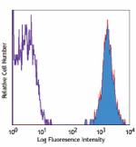
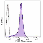
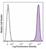
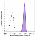
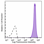
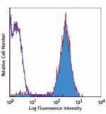
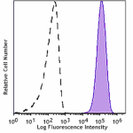
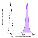
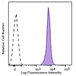
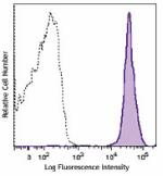
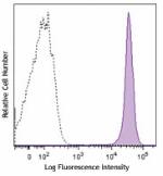
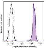
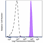
This data display is provided for general comparisons between formats.
Your actual data may vary due to variations in samples, target cells, instruments and their settings, staining conditions, and other factors.
If you need assistance with selecting the best format contact our expert technical support team.

Pricing & Availability
Clone
30-F11 (See other available formats)
Regulatory Status
RUO
Other Names
T200, Ly-5, LCA
Isotype
Rat IgG2b, κ
Ave. Rating
Submit a Review
Product Citations
publications
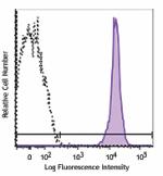
30-F11_Alx488_090507
C57BL/6 mouse splenocytes stained with 30-F11 Alexa Fluor® 488
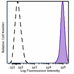
  • 30-F11_Alx488_090507
    C57BL/6 mouse splenocytes stained with 30-F11 Alexa Fluor® 488
  • 33_Mouse_Spleen_CD169_CD45
    Confocal image of C57BL/6 mouse spleen sample acquired using the IBEX method of highly multiplexed antibody-based imaging: CD169 (green) in Cycle 2 and CD45 (blue) in Cycle 3. Tissues were prepared using ~1% (vol/vol) formaldehyde and a detergent. Following fixation, samples are immersed in 30% (wt/vol) sucrose for cryoprotection. Images are courtesy of Drs. Andrea J. Radtke and Ronald N. Germain of the Center for Advanced Tissue Imaging (CAT-I) in the National Institute of Allergy and Infectious Diseases (NIAID, NIH).
Compare all formats See Alexa Fluor® 488 spectral data
Cat # Size Price Quantity Check Availability Save
103122 100 µg NOK1890
Check Availability


Need larger quantities of this item?
Request Bulk Quote
103121 25 µg NOK834
Check Availability


Need larger quantities of this item?
Request Bulk Quote
Description

CD45 is a 180-240 kD glycoprotein also known as the leukocyte common antigen (LCA), T200, or Ly-5. It is a member of the protein tyrosine phosphatase (PTP) family, expressed on all hematopoietic cells except mature erythrocytes and platelets. There are different isoforms of CD45 that arise from variable splicing of exons 4, 5, and 6, which encode A, B, and C determinants, respectively. CD45 plays a key role in TCR and BCR signal transduction. These isoforms are very specific to the activation and maturation state of the cell as well as cell type. The primary ligands for CD45 are galectin-1, CD2, CD3, CD4, TCR, CD22, and Thy-1.

Product Details
Technical Data Sheet (pdf)

Product Details

Verified Reactivity
Mouse
Antibody Type
Monoclonal
Host Species
Rat
Immunogen
Mouse thymus or spleen
Formulation
Phosphate-buffered solution, pH 7.2, containing 0.09% sodium azide.
Preparation
The antibody was purified by affinity chromatography and conjugated with Alexa Fluor® 488 under optimal conditions.
Concentration
0.5 mg/mL
Storage & Handling
The antibody solution should be stored undiluted between 2°C and 8°C, and protected from prolonged exposure to light. Do not freeze.
Application

FC - Quality tested

SB - Reported in the literature, not verified in house

Recommended Usage

Each lot of this antibody is quality control tested by immunofluorescent staining with flow cytometric analysis. For flow cytometric staining, the suggested use of this reagent is ≤ 0.25 µg per 106 cells in 100 µL volume. It is recommended that the reagent be titrated for optimal performance for each application.

* Alexa Fluor® 488 has a maximum emission of 519 nm when it is excited at 488 nm.


Alexa Fluor® and Pacific Blue™ are trademarks of Life Technologies Corporation.

View full statement regarding label licenses
Excitation Laser
Blue Laser (488 nm)
Application Notes

Clone 30-F11 reacts with all isoforms and both CD45.1 and CD45.2 alloantigens of CD45.

Additional reported applications (for relevant formats) include: immunoprecipitation3, complement-dependent cytotoxicity1,5, immunohistochemistry (acetone-fixed frozen sections, zinc-fixed paraffin-embedded sections and formalin-fixed paraffin-embedded sections)4,6, Western blotting7, and spatial biology (IBEX)10,11. The Ultra-LEAF™ purified antibody (Endotoxin < 0.01 EU/µg, Azide-Free, 0.2 µm filtered) is recommended for functional assays (Cat. No. 103163 and 103164).

Additional Product Notes

Iterative Bleaching Extended multi-pleXity (IBEX) is a fluorescent imaging technique capable of highly-multiplexed spatial analysis. The method relies on cyclical bleaching of panels of fluorescent antibodies in order to image and analyze many markers over multiple cycles of staining, imaging, and, bleaching. It is a community-developed open-access method developed by the Center for Advanced Tissue Imaging (CAT-I) in the National Institute of Allergy and Infectious Diseases (NIAID, NIH).

Application References
  1. Podd BS, et al. 2006. J. Immunol. 176:6532. (FC, CMCD) PubMed
  2. Haynes NM, et al. 2007. J. Immunol. 179:5099. (FC)
  3. Ledbetter JA, et al. 1979. Immunol. Rev. 47:63. (IP)
  4. Simon DI, et al. 2000. J. Clin. Invest. 105:293. (IHC)
  5. Seaman WE. 1983. J. Immunol. 130:1713. (CMCD)
  6. Cornet A, et al. 2001. P. Natl. Acad. Sci. USA 98:13306. (IHC)
  7. Tsuboi S and Fukuda M. 1998. J. Biol. Chem. 273:30680. (WB) PubMed
  8. Liu F, et al. 2012. Blood. 119:3295. PubMed
  9. Pelletier AN, et al. 2012. J. Immunol. 188:5561. PubMed
  10. Radtke AJ, et al. 2020. Proc Natl Acad Sci U S A. 117:33455-65. (SB) PubMed
  11. Radtke AJ, et al. 2022. Nat Protoc. 17:378-401. (SB) PubMed
Product Citations
  1. Preda MB, et al. 2021. Cell Death Dis. 12:566. PubMed
  2. Kuo PC, et al. 2021. Brain Commun. 3:fcab187. PubMed
  3. Wu Y, et al. 2021. Immunity. 54:2595. PubMed
  4. Wemlinger SM, et al. 2022. J Immunol. 208:1566. PubMed
  5. Liu H, et al. 2022. Cell Rep Med. 3:100660. PubMed
  6. Frederico B, et al. 2022. Dev Cell. 57:1957. PubMed
  7. Ullah I, et al. 2023. Cell Rep Med. 4:100893. PubMed
  8. Olivares-González L, et al. 2022. Antioxidants (Basel). 12: . PubMed
  9. Gilman KE, et al. 2023. Front Immunol. 13:1045710. PubMed
  10. Parida PK, et al. 2022. Cell Metab. 34:90. PubMed
  11. Gomez-Salinero JM, et al. 2022. Cell Stem Cell. 29:593. PubMed
  12. Luff SA, et al. 2022. Nat Cell Biol. 24:616. PubMed
  13. Battis K, et al. 2022. J Neurosci. 42:7673. PubMed
  14. Abdel-Haq R, et al. 2022. Elife. 11:. PubMed
  15. Axelrod ML, et al. 2022. Nature. 611:818. PubMed
  16. Gómez-Salinero JM, et al. 2022. Nat Cardiovasc Res. 1:882. PubMed
  17. Fan Z, et al. 2023. Angiogenesis. . PubMed
  18. Gilman KE, et al. 2023. Cancers (Basel). 15:. PubMed
  19. Palakurthi B, et al. 2023. Nat Commun. 14:2109. PubMed
  20. Fixsen BR, et al. 2023. Nat Immunol. . PubMed
  21. Marko C, et al. 2014. Invest Ophthalmol Vis Sci . 55:291. PubMed
  22. Giorgetti E, et al. 2020. Cell Reports. 29(6):1539-1554.e7.. PubMed
  23. Kazakevych J, et al. 2020. Genome Biol. 21:64. PubMed
  24. Sakai M, et al. 2020. Immunity. 51(4):655-670. PubMed
  25. Schmidleithner L et al. 2019. Immunity. 50(5):1232-1248 . PubMed
  26. Logan C, Bowen C, and Menko A 2017. Sci Rep. . 10.1038/s41598-017-16456-5. PubMed
  27. Petursdottir D, et al. 2017. Front Immunol. . 10.3389/fimmu.2017.01699. PubMed
  28. Nagai Y, et al. 2019. Front Immunol. 10:174. PubMed
  29. Khandelwal P, et al. 2013. PLoS One. 8:64193. PubMed
  30. Masschelein E, et al. 2020. Skelet Muscle. 10:21. PubMed
  31. Martínez-Sabadell A, et al. 2022. STAR Protoc. 3:101712. PubMed
  32. Rodriguez AB, et al. 2021. Cell Reports. 36(3):109422. PubMed
  33. Bornes L, et al. 2021. Life Sci Alliance. 4:00. PubMed
  34. Jenkins RW, et al. 2018. Cancer Discov. 8:196. PubMed
  35. Dubey LK, et al. 2019. Cell Rep. 27:2442. PubMed
  36. DeDreu J, et al. 2020. FASEB J. 34:9316. PubMed
  37. Dos Santos Dias L, et al. 2021. PLoS Pathog. e1009324:17. PubMed
  38. Hallett JM, et al. 2022. Cell Stem Cell. 29:355. PubMed
  39. Mehta AK, et al. 2021. Nat Cancer. 2:66. PubMed
  40. Yip HYK, et al. 2021. STAR Protocols. 2(3):100765. PubMed
  41. Chen I, et al. 2016. Sci Rep. 6:27195. PubMed
  42. Duarte J, et al. 2016. J Immunol. 197(12):4838-4847. PubMed
  43. Jung Y, et al. 2021. Nat Commun. 12:3872. PubMed
  44. Scarneo SA, et al. 2022. Sci Rep. 12:18091. PubMed
  45. Gutknecht M, et al. 2017. Nat Commun. 10.1038/s41467-017-00488-6. PubMed
  46. Ferrari G, et al. 2011. Invest Ophthalmol Vis Sci. 52:2532. PubMed
  47. Mizbani A, et al. 2016. Development. 143: 4137 - 4148. PubMed
  48. Burns JC, et al. 2020. eLife. 9:00. PubMed
  49. Wara AK, et al. 2020. Cell Rep. 33:108550. PubMed
  50. Wu H, et al. 2021. Cell Death Discov. 7:225. PubMed
  51. Lustgarten Guahmich N, et al. 2020. Angiogenesis. 23:443. PubMed
  52. Limbad C, et al. 2022. iScience. 25:103848. PubMed
  53. Yip HYK, et al. 2020. Molecular Cell. 80(2):279-295.e8. PubMed
  54. Liu X, et al. 2020. Cell Host Microbe. 28(5):683-698.e6. PubMed
  55. Dobson HE, et al. 2020. Mucosal Immunol. 0.901388889. PubMed
  56. Biton M et al. 2018. Cell. 175(5):1307-1320 . PubMed
  57. Alam Z, et al. 2020. Cell Rep. 107825:31. PubMed
  58. Shibad V, et al. 2021. Front Immunol. 12:722451. PubMed
  59. Wüthrich M, et al. 2021. MBio. 12:e0201821. PubMed
  60. Li D, et al. 2022. Commun Biol. 5:271. PubMed
  61. Braun D, et al. 2013. J Biol Chem. 288:2689. PubMed
  62. Pang M, et al. 2009. J Immunol. 182:7001. PubMed
  63. Wu X, et al. 2018. Cell. 172:423. PubMed
  64. von Roemeling CA, et al. 2020. Nat Commun. 11:1508. PubMed
  65. Ding H, et al. 2016. Nat Commun. 7:11533. PubMed
  66. Franklin DA, et al. 2020. JCI Insight. 5:. PubMed
  67. Steele NG, et al. 2021. Clin Cancer Res. 27:2023. PubMed
  68. Kazakevych J, et al. 2019. Sci Rep. 9:10410. PubMed
  69. Peng W, et al. 2018. Mol Cell Biol. 38:e00427. PubMed
  70. Kuo P, et al. 2016. J Am Heart Assoc. 5: 002610. PubMed
  71. Markworth JF, et al. 2020. JCI Insight. 5:00. PubMed
  72. Reed-Geaghan EG, et al. 2020. J Exp Med. 217:00:00. PubMed
  73. Pantelidou C, et al. 2022. NPJ Breast Cancer. 8:102. PubMed
  74. Lasch M, et al. 2020. Front Physiol. 11:576736. PubMed
  75. DeDreu J, et al. 2022. FASEB J. 36:e21995. PubMed
  76. Li Q, et al. 2020. Am J Pathol. 190:2453. PubMed
  77. Lee EKS, et al. 2018. Cell Host Microbe. 23:121. PubMed
  78. Wang Y, et al. 2019. Front Cell Infect Microbiol. 9:286. PubMed
  79. Ye Y, et al. 2011. Am J Physiol Lung Cell Mol Physiol. 300:L216. PubMed
  80. RL M, et al. 2015. Proc Natl Acad Sci U S A. 112:6506-6514. PubMed
  81. Tian F, et al. 2016. Nat Commun. 7:13283. PubMed
  82. Yang FM, et al. 2022. Front Immunol. 13:918241. PubMed
  83. Lal JC, et al. 2021. Breast Cancer Res. 23:83. PubMed
  84. Xiang W, et al. 2020. Signal Transduct Target Ther. 0.374305556. PubMed
  85. Denk F, et al. 2016. Cell Rep. 15: 1771-1781. PubMed
  86. Qi S, et al. 2020. Theranostics. 10:1814. PubMed
  87. Preda MB, et al. 2021. Cell Death Dis. 12:566. PubMed
  88. Kuo PC, et al. 2021. Brain Commun. 3:fcab187. PubMed
  89. Wu Y, et al. 2021. Immunity. 54:2595. PubMed
  90. Wemlinger SM, et al. 2022. J Immunol. 208:1566. PubMed
  91. Liu H, et al. 2022. Cell Rep Med. 3:100660. PubMed
  92. Frederico B, et al. 2022. Dev Cell. 57:1957. PubMed
  93. Ullah I, et al. 2023. Cell Rep Med. 4:100893. PubMed
  94. Olivares-González L, et al. 2022. Antioxidants (Basel). 12: . PubMed
  95. Gilman KE, et al. 2023. Front Immunol. 13:1045710. PubMed
  96. Parida PK, et al. 2022. Cell Metab. 34:90. PubMed
  97. Gomez-Salinero JM, et al. 2022. Cell Stem Cell. 29:593. PubMed
  98. Luff SA, et al. 2022. Nat Cell Biol. 24:616. PubMed
  99. Battis K, et al. 2022. J Neurosci. 42:7673. PubMed
  100. Abdel-Haq R, et al. 2022. Elife. 11:. PubMed
  101. Axelrod ML, et al. 2022. Nature. 611:818. PubMed
  102. Gómez-Salinero JM, et al. 2022. Nat Cardiovasc Res. 1:882. PubMed
  103. Fan Z, et al. 2023. Angiogenesis. . PubMed
  104. Gilman KE, et al. 2023. Cancers (Basel). 15:. PubMed
  105. Palakurthi B, et al. 2023. Nat Commun. 14:2109. PubMed
  106. Fixsen BR, et al. 2023. Nat Immunol. . PubMed
  107. Marko C, et al. 2014. Invest Ophthalmol Vis Sci . 55:291. PubMed
  108. Giorgetti E, et al. 2020. Cell Reports. 29(6):1539-1554.e7.. PubMed
  109. Kazakevych J, et al. 2020. Genome Biol. 21:64. PubMed
  110. Sakai M, et al. 2020. Immunity. 51(4):655-670. PubMed
  111. Schmidleithner L et al. 2019. Immunity. 50(5):1232-1248 . PubMed
  112. Logan C, Bowen C, and Menko A 2017. Sci Rep. . 10.1038/s41598-017-16456-5. PubMed
  113. Petursdottir D, et al. 2017. Front Immunol. . 10.3389/fimmu.2017.01699. PubMed
  114. Nagai Y, et al. 2019. Front Immunol. 10:174. PubMed
  115. Khandelwal P, et al. 2013. PLoS One. 8:64193. PubMed
  116. Masschelein E, et al. 2020. Skelet Muscle. 10:21. PubMed
  117. Martínez-Sabadell A, et al. 2022. STAR Protoc. 3:101712. PubMed
  118. Rodriguez AB, et al. 2021. Cell Reports. 36(3):109422. PubMed
  119. Bornes L, et al. 2021. Life Sci Alliance. 4:00. PubMed
  120. Jenkins RW, et al. 2018. Cancer Discov. 8:196. PubMed
  121. Dubey LK, et al. 2019. Cell Rep. 27:2442. PubMed
  122. DeDreu J, et al. 2020. FASEB J. 34:9316. PubMed
  123. Dos Santos Dias L, et al. 2021. PLoS Pathog. e1009324:17. PubMed
  124. Hallett JM, et al. 2022. Cell Stem Cell. 29:355. PubMed
  125. Mehta AK, et al. 2021. Nat Cancer. 2:66. PubMed
  126. Yip HYK, et al. 2021. STAR Protocols. 2(3):100765. PubMed
  127. Chen I, et al. 2016. Sci Rep. 6:27195. PubMed
  128. Duarte J, et al. 2016. J Immunol. 197(12):4838-4847. PubMed
  129. Jung Y, et al. 2021. Nat Commun. 12:3872. PubMed
  130. Scarneo SA, et al. 2022. Sci Rep. 12:18091. PubMed
  131. Gutknecht M, et al. 2017. Nat Commun. 10.1038/s41467-017-00488-6. PubMed
  132. Ferrari G, et al. 2011. Invest Ophthalmol Vis Sci. 52:2532. PubMed
  133. Mizbani A, et al. 2016. Development. 143: 4137 - 4148. PubMed
  134. Burns JC, et al. 2020. eLife. 9:00. PubMed
  135. Wara AK, et al. 2020. Cell Rep. 33:108550. PubMed
  136. Wu H, et al. 2021. Cell Death Discov. 7:225. PubMed
  137. Lustgarten Guahmich N, et al. 2020. Angiogenesis. 23:443. PubMed
  138. Limbad C, et al. 2022. iScience. 25:103848. PubMed
  139. Yip HYK, et al. 2020. Molecular Cell. 80(2):279-295.e8. PubMed
  140. Liu X, et al. 2020. Cell Host Microbe. 28(5):683-698.e6. PubMed
  141. Dobson HE, et al. 2020. Mucosal Immunol. 0.901388889. PubMed
  142. Biton M et al. 2018. Cell. 175(5):1307-1320 . PubMed
  143. Alam Z, et al. 2020. Cell Rep. 107825:31. PubMed
  144. Shibad V, et al. 2021. Front Immunol. 12:722451. PubMed
  145. Wüthrich M, et al. 2021. MBio. 12:e0201821. PubMed
  146. Li D, et al. 2022. Commun Biol. 5:271. PubMed
  147. Braun D, et al. 2013. J Biol Chem. 288:2689. PubMed
  148. Pang M, et al. 2009. J Immunol. 182:7001. PubMed
  149. Wu X, et al. 2018. Cell. 172:423. PubMed
  150. von Roemeling CA, et al. 2020. Nat Commun. 11:1508. PubMed
  151. Ding H, et al. 2016. Nat Commun. 7:11533. PubMed
  152. Franklin DA, et al. 2020. JCI Insight. 5:. PubMed
  153. Steele NG, et al. 2021. Clin Cancer Res. 27:2023. PubMed
  154. Kazakevych J, et al. 2019. Sci Rep. 9:10410. PubMed
  155. Peng W, et al. 2018. Mol Cell Biol. 38:e00427. PubMed
  156. Kuo P, et al. 2016. J Am Heart Assoc. 5: 002610. PubMed
  157. Markworth JF, et al. 2020. JCI Insight. 5:00. PubMed
  158. Reed-Geaghan EG, et al. 2020. J Exp Med. 217:00:00. PubMed
  159. Pantelidou C, et al. 2022. NPJ Breast Cancer. 8:102. PubMed
  160. Lasch M, et al. 2020. Front Physiol. 11:576736. PubMed
  161. DeDreu J, et al. 2022. FASEB J. 36:e21995. PubMed
  162. Li Q, et al. 2020. Am J Pathol. 190:2453. PubMed
  163. Lee EKS, et al. 2018. Cell Host Microbe. 23:121. PubMed
  164. Wang Y, et al. 2019. Front Cell Infect Microbiol. 9:286. PubMed
  165. Ye Y, et al. 2011. Am J Physiol Lung Cell Mol Physiol. 300:L216. PubMed
  166. RL M, et al. 2015. Proc Natl Acad Sci U S A. 112:6506-6514. PubMed
  167. Tian F, et al. 2016. Nat Commun. 7:13283. PubMed
  168. Yang FM, et al. 2022. Front Immunol. 13:918241. PubMed
  169. Lal JC, et al. 2021. Breast Cancer Res. 23:83. PubMed
  170. Xiang W, et al. 2020. Signal Transduct Target Ther. 0.374305556. PubMed
  171. Denk F, et al. 2016. Cell Rep. 15: 1771-1781. PubMed
  172. Qi S, et al. 2020. Theranostics. 10:1814. PubMed
RRID
AB_493531 (BioLegend Cat. No. 103122)
AB_493532 (BioLegend Cat. No. 103121)

Antigen Details

Structure
Protein tyrosine phosphatase (PTP) family, 180-240 kD
Distribution

All hematopoietic cells except mature erythrocytes and platelets

Function
Phosphatase, T and B cell activation
Ligand/Receptor
Galectin-1, CD2, CD3, CD4, TCR, CD22, Thy-1
Cell Type
B cells, Dendritic cells, Mesenchymal Stem Cells, Tregs
Biology Area
Cell Biology, Immunology, Inhibitory Molecules, Innate Immunity, Neuroscience, Neuroscience Cell Markers, Stem Cells
Molecular Family
CD Molecules
Antigen References

1. Barclay A, et al. 1997. The Leukocyte Antigen FactsBook Academic Press.
2. Trowbridge IS, et al. 1993. Annu. Rev. Immunol. 12:85.
3. Kishihara K, et al. 1993. Cell 74:143.
4. Pulido R, et al. 1988. J. Immunol. 140:3851.

Gene ID
19264 View all products for this Gene ID
UniProt
View information about CD45 on UniProt.org

Related FAQs

If an antibody clone has been previously successfully used in IBEX in one fluorescent format, will other antibody formats work as well?

It’s likely that other fluorophore conjugates to the same antibody clone will also be compatible with IBEX using the same sample fixation procedure. Ultimately a directly conjugated antibody’s utility in fluorescent imaging and IBEX may be specific to the sample and microscope being used in the experiment. Some antibody clone conjugates may perform better than others due to performance differences in non-specific binding, fluorophore brightness, and other biochemical properties unique to that conjugate.

Will antibodies my lab is already using for fluorescent or chromogenic IHC work in IBEX?

Fundamentally, IBEX as a technique that works much in the same way as single antibody panels or single marker IF/IHC. If you’re already successfully using an antibody clone on a sample of interest, it is likely that clone will have utility in IBEX. It is expected some optimization and testing of different antibody fluorophore conjugates will be required to find a suitable format; however, legacy microscopy techniques like chromogenic IHC on fixed or frozen tissue is an excellent place to start looking for useful antibodies.

Are other fluorophores compatible with IBEX?

Over 18 fluorescent formats have been screened for use in IBEX, however, it is likely that other fluorophores are able to be rapidly bleached in IBEX. If a fluorophore format is already suitable for your imaging platform it can be tested for compatibility in IBEX.

The same antibody works in one tissue type but not another. What is happening?

Differences in tissue properties may impact both the ability of an antibody to bind its target specifically and impact the ability of a specific fluorophore conjugate to overcome the background fluorescent signal in a given tissue. Secondary stains, as well as testing multiple fluorescent conjugates of the same clone, may help to troubleshoot challenging targets or tissues. Using a reference control tissue may also give confidence in the specificity of your staining.

How can I be sure the staining I’m seeing in my tissue is real?

In general, best practices for validating an antibody in traditional chromogenic or fluorescent IHC are applicable to IBEX. Please reference the Nature Methods review on antibody based multiplexed imaging for resources on validating antibodies for IBEX.

Other Formats

View All CD45 Reagents Request Custom Conjugation
Description Clone Applications
PE anti-mouse CD45 30-F11 FC
PE/Cyanine5 anti-mouse CD45 30-F11 FC
Purified anti-mouse CD45 30-F11 FC,IHC-F,CyTOF®,IP,CMCD,IHC,WB
PE/Cyanine7 anti-mouse CD45 30-F11 FC
PE/Dazzle™ 594 anti-mouse CD45 30-F11 FC
Brilliant Violet 711™ anti-mouse CD45 30-F11 FC
Brilliant Violet 785™ anti-mouse CD45 30-F11 FC
Brilliant Violet 650™ anti-mouse CD45 30-F11 FC
APC/Fire™ 750 anti-mouse CD45 30-F11 FC
Brilliant Violet 750™ anti-mouse CD45 30-F11 FC
TotalSeq™-A0096 anti-mouse CD45 30-F11 PG
TotalSeq™-B0096 anti-mouse CD45 30-F11 PG
Ultra-LEAF™ Purified anti-mouse CD45 30-F11 FC,CyTOF®,IP,CMCD,IHC,WB
Spark Blue™ 550 anti-mouse CD45 30-F11 FC
Spark NIR™ 685 anti-mouse CD45 30-F11 FC
TotalSeq™-C0096 anti-mouse CD45 30-F11 PG
Spark YG™ 570 anti-mouse CD45 30-F11 IHC-F
PE/Fire™ 640 anti-mouse CD45 30-F11 FC
APC/Fire™ 810 anti-mouse CD45 30-F11 FC
PE/Fire™ 700 anti-mouse CD45 30-F11 FC
APC anti-mouse CD45 30-F11 FC
Biotin anti-mouse CD45 30-F11 FC
FITC anti-mouse CD45 30-F11 FC
APC/Cyanine7 anti-mouse CD45 30-F11 FC
Alexa Fluor® 488 anti-mouse CD45 30-F11 FC,SB
Alexa Fluor® 647 anti-mouse CD45 30-F11 FC,ICC,IHC,3D IHC,SB
Pacific Blue™ anti-mouse CD45 30-F11 FC
Alexa Fluor® 700 anti-mouse CD45 30-F11 FC,SB
PerCP/Cyanine5.5 anti-mouse CD45 30-F11 FC
PerCP anti-mouse CD45 30-F11 FC
Alexa Fluor® 594 anti-mouse CD45 30-F11 IHC-F,FC,3D IHC
Brilliant Violet 421™ anti-mouse CD45 30-F11 FC,SB
Brilliant Violet 570™ anti-mouse CD45 30-F11 FC
Brilliant Violet 510™ anti-mouse CD45 30-F11 FC
Brilliant Violet 605™ anti-mouse CD45 30-F11 FC
Purified anti-mouse CD45 (Maxpar® Ready) 30-F11 FC,CyTOF®
Spark Violet™ 538 anti-mouse CD45 30-F11 FC
Spark YG™ 593 anti-mouse CD45 30-F11 FC
Spark Blue™ 574 anti-mouse CD45 Antibody 30-F11 FC
Spark Blue™ 515 anti-mouse CD45 30-F11 FC
Spark UV™ 387 anti-mouse CD45 30-F11 FC
PE/Fire™ 810 anti-mouse CD45 30-F11 FC
Spark PLUS UV395™ anti-mouse CD45 30-F11 FC
Spark Red™ 718 anti-mouse CD45 30-F11 FC
Spark YG™ 581 anti-mouse CD45 (Flexi-Fluor™) 30-F11 FC
Spark PLUS V475™ anti-mouse CD45 Antibody 30-F11 FC
StarBright UltraViolet 740 anti-mouse CD45 30-F11 FC
Go To Top Version: 5    Revision Date: 04.20.2022

For Research Use Only. Not for diagnostic or therapeutic use.


This product is supplied subject to the terms and conditions, including the limited license, located at www.biolegend.com/terms) ("Terms") and may be used only as provided in the Terms. Without limiting the foregoing, BioLegend products may not be used for any Commercial Purpose as defined in the Terms, resold in any form, used in manufacturing, or reverse engineered, sequenced, or otherwise studied or used to learn its design or composition without express written approval of BioLegend. Regardless of the information given in this document, user is solely responsible for determining any license requirements necessary for user’s intended use and assumes all risk and liability arising from use of the product. BioLegend is not responsible for patent infringement or any other risks or liabilities whatsoever resulting from the use of its products.


BioLegend, the BioLegend logo, and all other trademarks are property of BioLegend, Inc. or their respective owners, and all rights are reserved.


8999 BioLegend Way, San Diego, CA 92121 www.biolegend.com
Toll-Free Phone: 1-877-Bio-Legend (246-5343) Phone: (858) 768-5800 Fax: (877) 455-9587

This data display is provided for general comparisons between formats.
Your actual data may vary due to variations in samples, target cells, instruments and their settings, staining conditions, and other factors.
If you need assistance with selecting the best format contact our expert technical support team.

ProductsHere

Login / Register
Remember me
Forgot your password? Reset password?
Create an Account